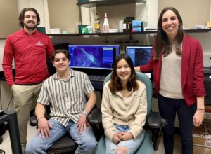
Starting a neuroscience lab with Dr. Michelle Bedenbaugh
We caught up with Dr. Bedenbaugh about her experience as a new investigator, what starting a new lab entails, and why she chose to bring LifeCanvas with her on her next big scientific journey.

We caught up with Dr. Bedenbaugh about her experience as a new investigator, what starting a new lab entails, and why she chose to bring LifeCanvas with her on her next big scientific journey.
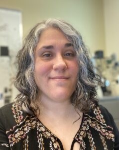
Dr. Montero Llopis shares her insights on the MicRoN Core’s vision, its adoption of LifeCanvas technologies, and how our values align to support scientific advancement.
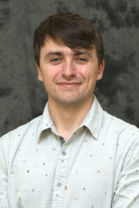
Dr. Andrew Stone shares his insights on SmartBatch+ and SmartSPIM and how they would be a great addition to the core.

Dr. Tanya Daigle used LifeCanvas’ services to acquire 3D images of spinal motor neurons illuminated by enhancer AAV.
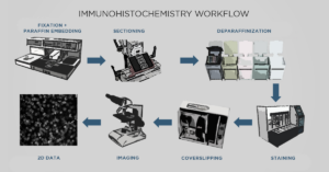
Immunohistochemistry (IHC) and 3D histology are different approaches to detecting specific proteins in biological tissue.
Dr. Sam Centanni uses the LifeCanvas pipeline to investigate the neurobiological overlap between stress and predisposition to alcohol abuse, as well as long-term effects of alcohol on the brain.