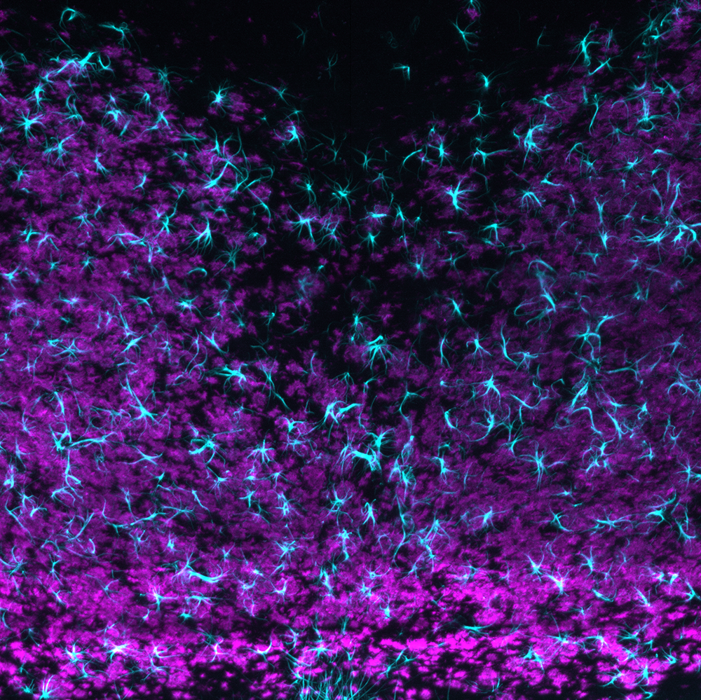
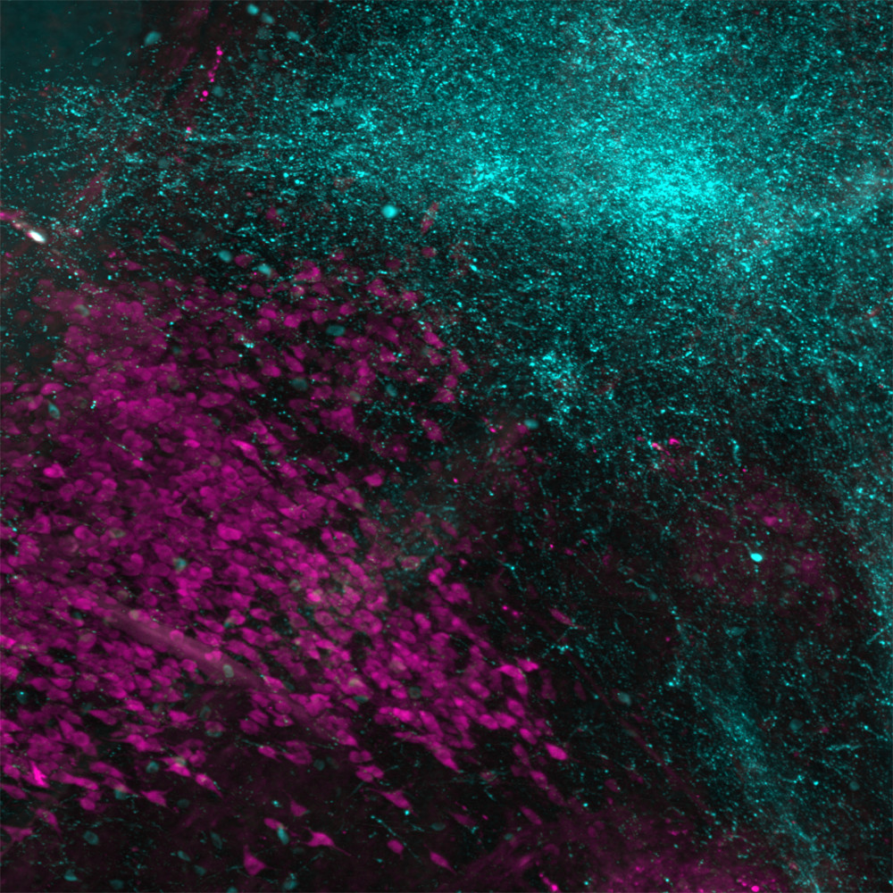
Dr. Sam Centanni uses the LifeCanvas pipeline to investigate the neurobiological overlap between stress and predisposition to alcohol abuse, as well as long-term effects of alcohol on the brain.


Dr. Sam Centanni uses the LifeCanvas pipeline to investigate the neurobiological overlap between stress and predisposition to alcohol abuse, as well as long-term effects of alcohol on the brain.

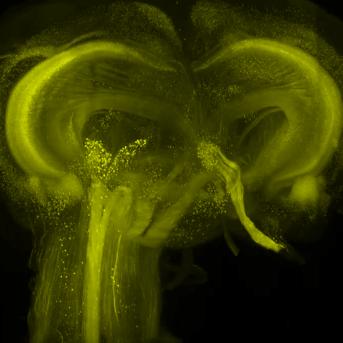
Dr. Mike Taormina uses SmartSPIM light sheet microscopy to rapidly image whole mouse brains, streamlining workflows at the Allen Institute.

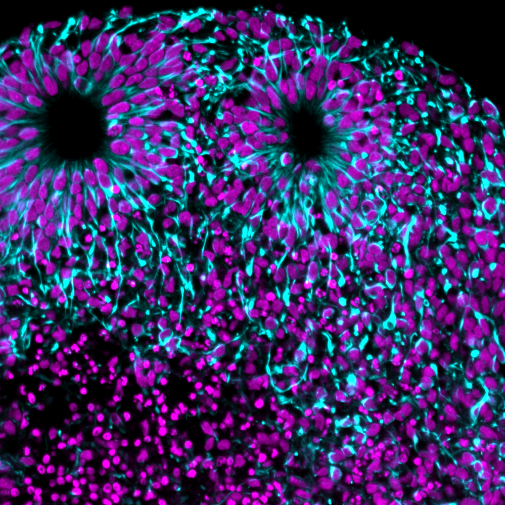
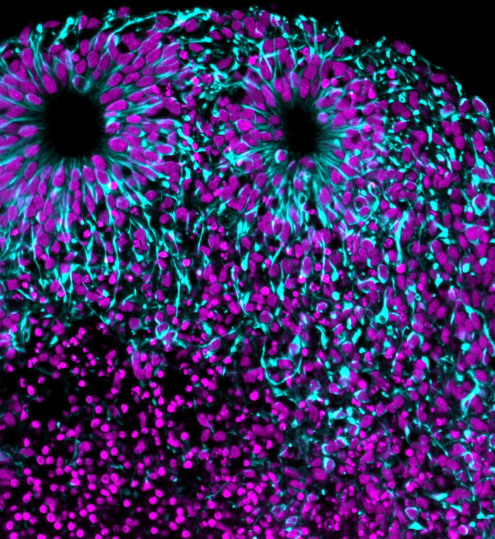
Dr. Kim's lab at Penn State uses the SmartSPIM light sheet microscope to map and create 3D atlases of the developing mouse brain.

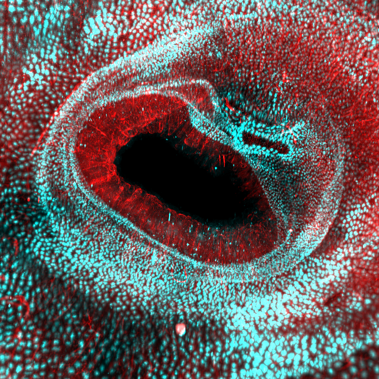
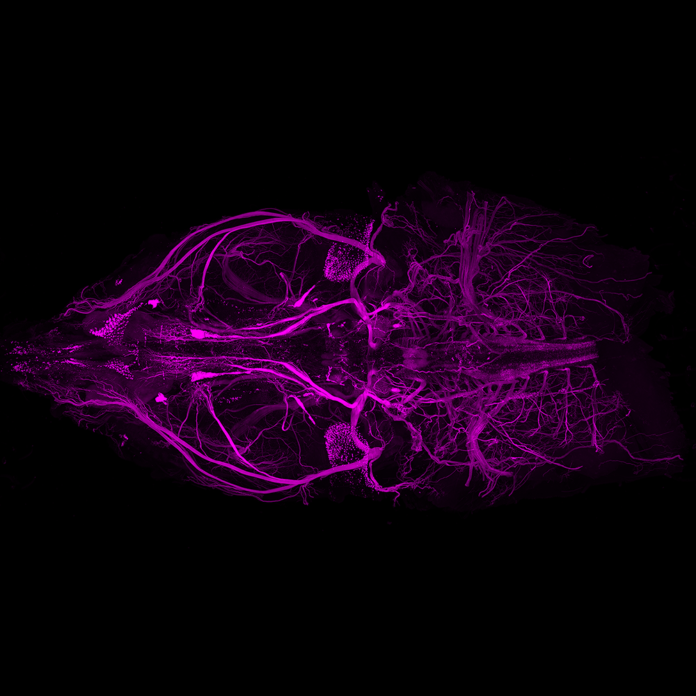
Dr. Ye's lab at the Scripps Research Institute uses SmartSPIM to illuminate interactions between the central and peripheral nervous systems.