Dr. Wenqiang Chen leverages LifeCanvas whole brain mapping to study the interaction of insulin resistance with Alzheimer's disease.

Depositions of aberrant protein aggregates in the brain are recognized hallmarks of major neurodegenerative disorders like Alzheimer’s, Parkinson’s, and Huntington’s diseases. We can process and analyze samples for β-amyloid deposition (e.g. in Alzheimer’s), as well as for the accumulation of α-synuclein in Lewy Bodies (e.g. in Parkinson’s).
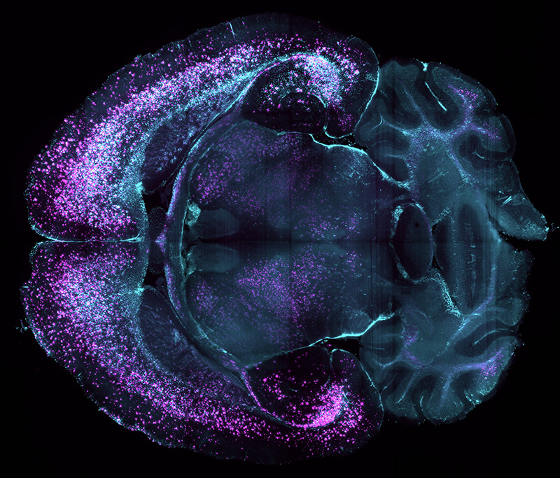
Localized damage to diseased brain parenchyma is often accompanied by a neuroinflammatory response, such as activation and recruitment of reactive glia (e.g. astrocytes, microglia). Our technologies can be used to assess these responses through mapping of astrocytes (GFAP+) and microglia (IBA1+).
Pictued above: Astrocytes marked by GFAP (magenta) cluster around β-amyloid plaques (cyan), in brain of 7-month-old homozygous ARTE10 male mouse from Taconic Biosciences, Alzheimer’s disease model #16347.
5xFAD mouse brain model of Alzheimer’s disease. Sample courtesy of Drs. David Elliott and Jonathan Epp, University of Calgary.
Upon receiving your samples, LifeCanvas technicians:
You send us: PFA-fixed brain samples from your disease model animals
We deliver:
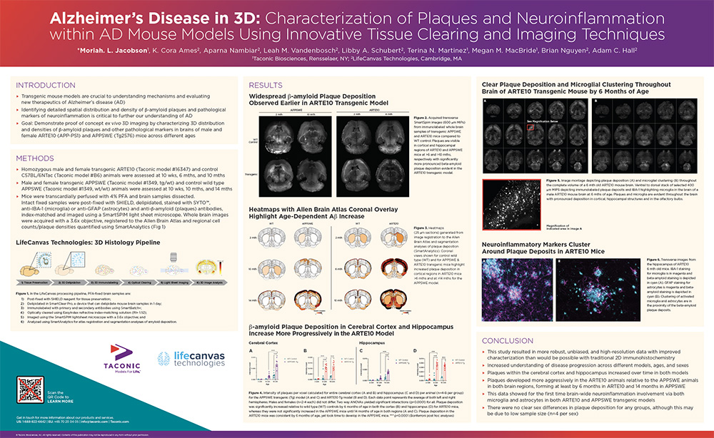
Poster created in collaboration with Taconic Biosciences for the Society for Neuroscience’s 2022 conference. Click to download PDF.
Dr. Wenqiang Chen leverages LifeCanvas whole brain mapping to study the interaction of insulin resistance with Alzheimer's disease.
What makes the mouse model so popular for disease research? We discuss the benefits, complications, and ongoing improvements of murine...
3D tissue clearing, labeling, imaging, and analysis tools provide critical spatial data for investigating neurological disorders.
Dr. Jonathan Epp's lab at the University of Calgary uses LifeCanvas tools to robustly characterize mouse models for Alzheimer's.