
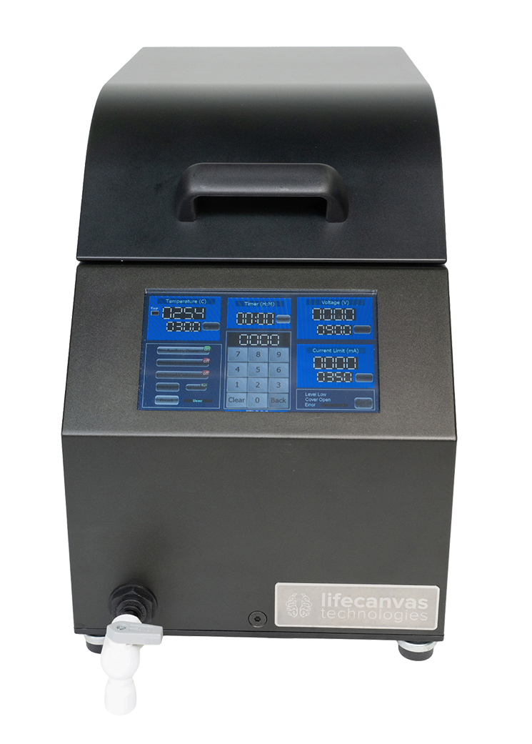
Our easy-to-use system combines active tissue clearing and immunolabeling into one high-throughput device, allowing you to switch between clearing and labeling modes simply by changing buffers. SmartBatch+ is compatible with different tissue types and large intact samples.
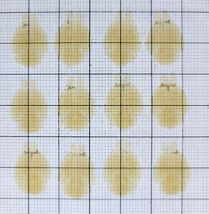
An advance on our CLARITY tissue clearing method, Clear+ provides maximum optical transparency with no tissue expansion or contraction. In immunolabeling mode, eFLASH and patented SE technologies uniformly label whole organs. Unlike other methods like iDISCO and CUBIC, SmartBatch+ actively preserves fluorescent protein signal.
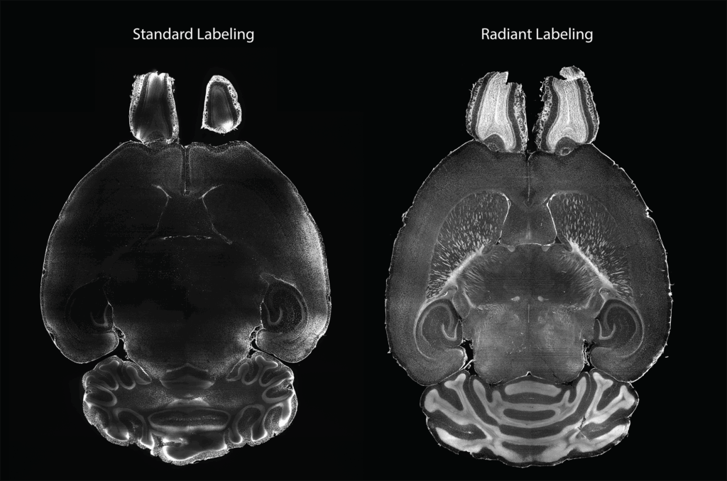
Radiant is a new primary labeling buffer system designed to improve antibody binding efficiency and accuracy in targets that are not well covered by standard methods. By modulating binding affinity, Radiant allows some antibodies to distribute more evenly throughout the sample, ensuring better coverage in cases like:
Note: All new SmartBatch+ devices come with Radiant hardware pre-installed, and are still fully compatible with the standard buffer system. Radiant is not intended to replace the Standard buffer system for all targets – please check the validated antibody list to see which system is best for your target.
Already have a SmartBatch+? Contact us to learn more.
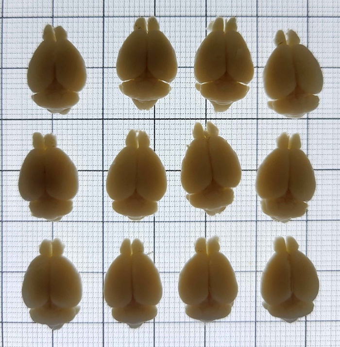
SmartBatch+ actively clears and labels up to 12 whole mouse brains or comparably sized samples in as little as one day, and 2 whole rat brains in just 3-4 days! This provides unparalleled consistency for multiple treatment groups. Its small footprint also allows ample space for multiple devices.
SmartBatch+ combined with SHIELD tissue preservation protects fluorescent protein signal, antigenicity, and molecular and physical architecture through all tissue processing steps. Clear+ is currently the only tissue clearing technique that preserves sample morphology. This ensures your samples will produce information-rich images.
Use just 4-20 μg of antibody per target to label a sample the size of a whole mouse brain — up to an order of magnitude less than passive staining. Save hundreds of dollars per sample per antibody, in primary antibody alone! SmartBatch+’s turnkey design is also labor-efficient, requiring <12 mins hands-on time per sample to clear and label.
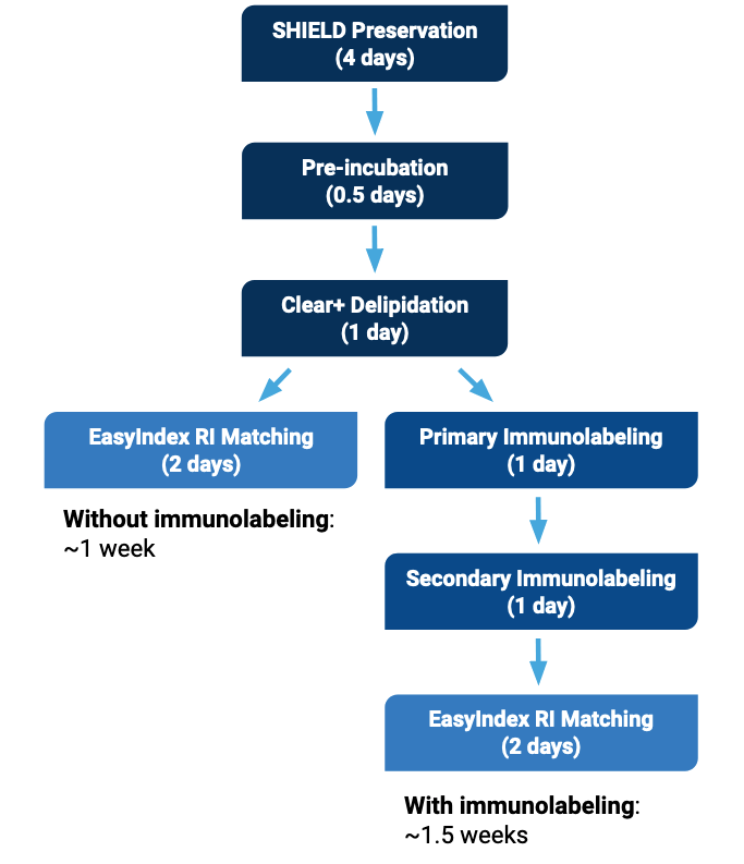
Our full pipeline kits include all reagents and buffers to protect, delipidate, label and index-match your tissue samples with SmartBatch+.
Reagent Kit for Full Pipeline for 25 Mouse Brain Sized Samples
Reagent Kit for Full Pipeline for 50 Mouse Brain Sized Samples
Reagent Kit for Full Pipeline for 100 Mouse Brain Sized Samples
Mohammed A. Khallaf, Daniel W. Hart, Wenhan Luo, Firdevs Murad, Felipe Cybis Pereira, Daniel Mendez-Aranda, Nicole Hagenah, Alice Rossi, Valérie Bégay, Jan Okrouhlík, Dietmar Krautwurst, Mungo Kisinza Ngalameno, Andre Ganswindt, Alison J. Barker, Radim Šumbera, Markus Knaden, Sophie Pezet, Andrew Woehler, Bill S. Hansson, Nigel C. Bennett & Gary R. Lewin
Tatiyana L. Adkins, Amanda L. Salazar, Jincy R. Little, Ellen C. Howard, Samuel W. Centanni
Shreyas M. Suryanarayana, Xu An, Yongjun Qian, Shengli Zhao, Hemanth Mohan, Z. Josh Huang