
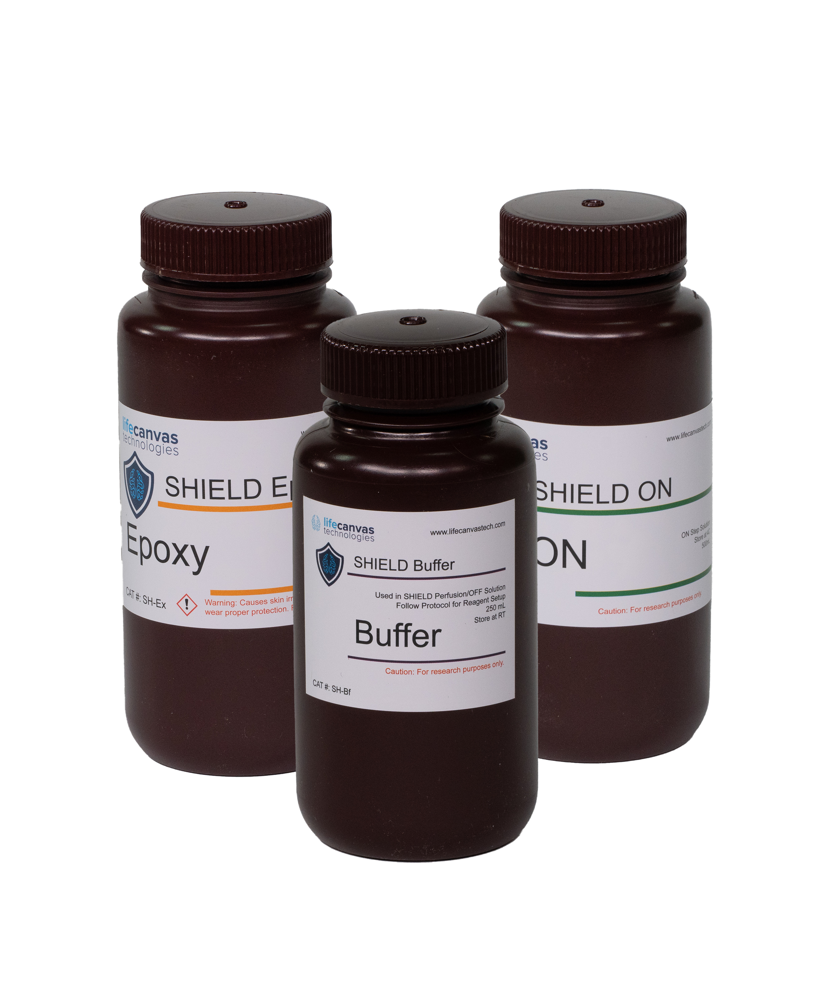
Our novel SHIELD tissue preservation technique forms intramolecular bonds using polyfunctional, flexible epoxides to stabilize tissue architecture and safeguard the sample’s endogenous fluorescence, protein antigenicity and nucleic acids. SHIELD avoids the variability of hydrogel embedding and the information loss from PFA preservation, protecting specimens for multiple rounds of processing.
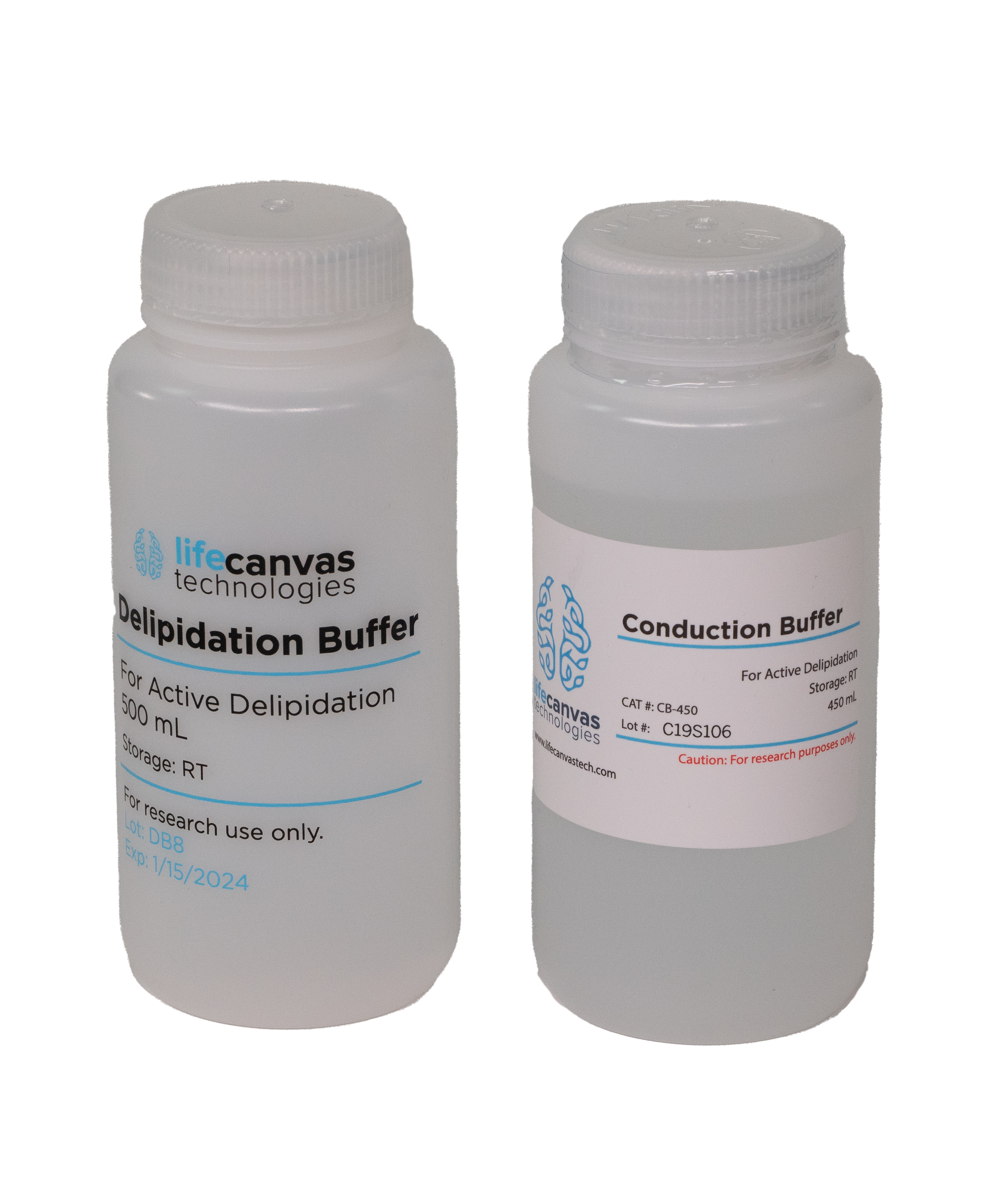
Our new Clear+ tissue clearing technique is the only method that delipidates samples with no change in morphology and with minimal impact on structural integrity. Fully delipidate whole mouse brains or comparably sized samples in just one day with SmartBatch+, or in one week with our passive clearing kit.
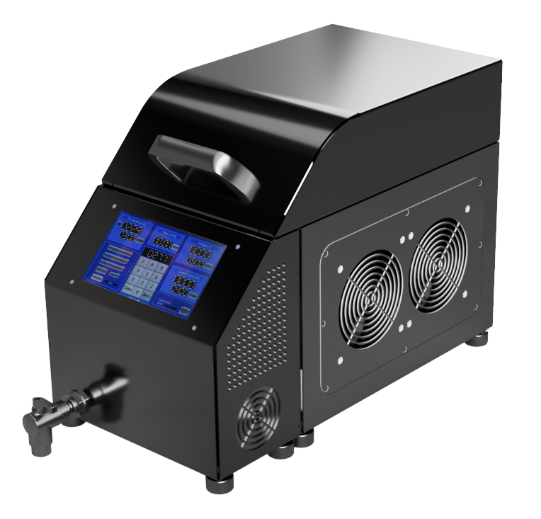
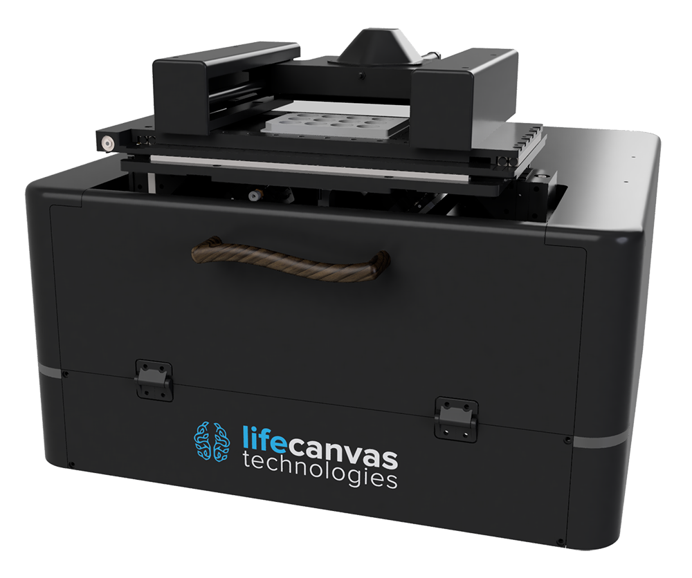
Our turn-key SmartBatch+ system combines electrophoretic tissue clearing and immunolabeling into one high-throughput device. Leverage the Clear+ tissue clearing method, along with eFLASH and patented stochastic electrotransport technologies, to rapidly clear and label whole organs. Key highlights and features include:
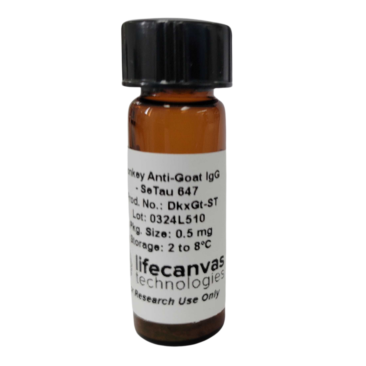
Our SeTau-647 Secondary Antibody is the only commercially available pre-conjugated SeTau Secondary that will provide superior photostability when labeling. Ideal for light sheet, 2P Imaging, expansion tissues, super-resolution, or live cell imaging where fluorescence stability is paramount.
Specifications:
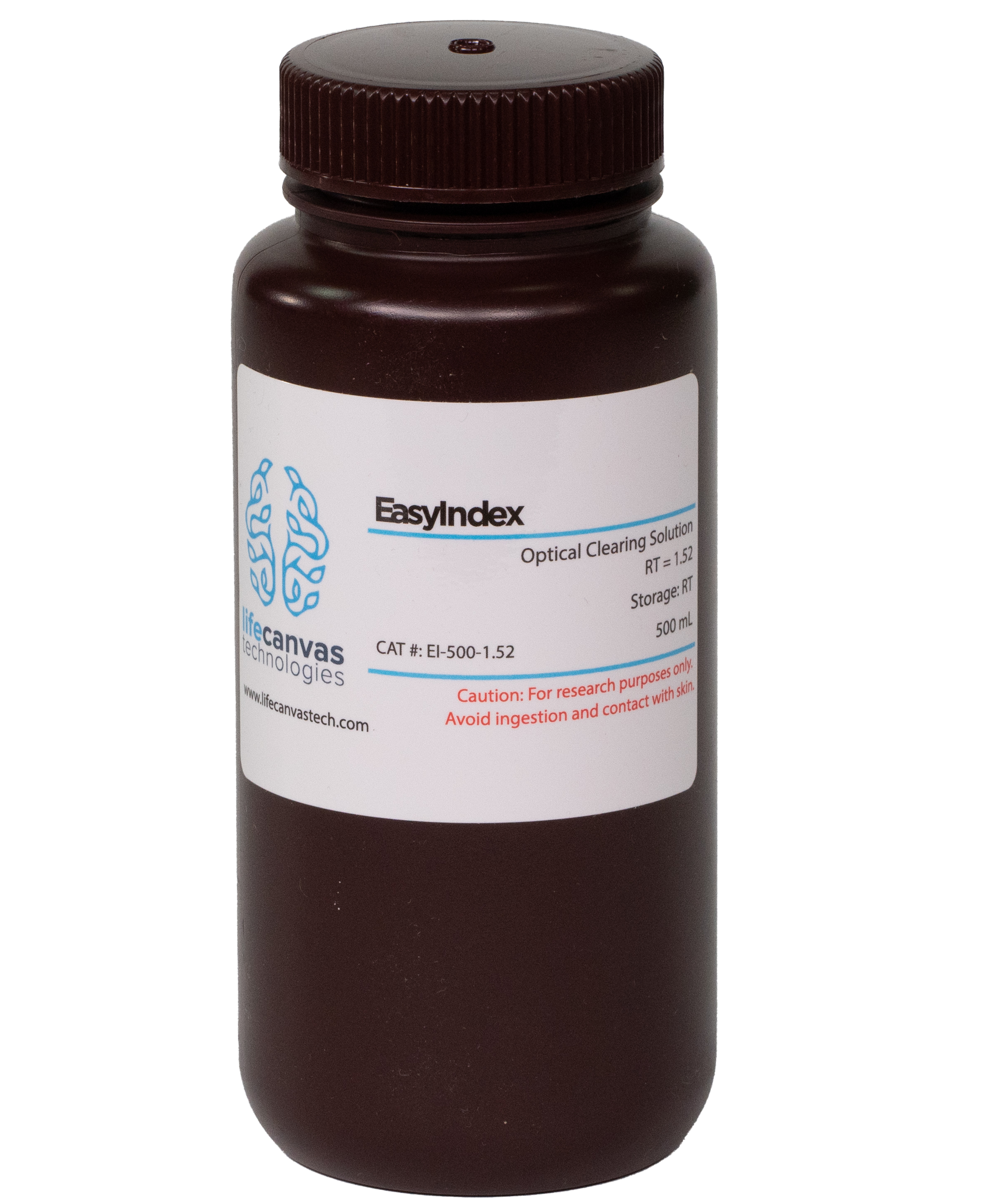
Our preformulated EasyIndex solution raises and homogenize the refractive index of delipidated tissue samples, rendering them fully transparent. This enables light penetration into the sample and ensures the acquisition of high-resolution, in-focus image data.
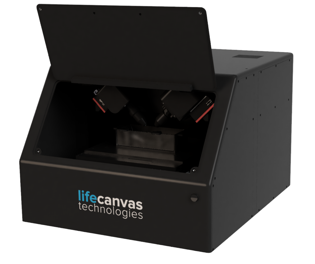
DALISPIM is an open-top, inverted light sheet microscope that combines high-speed, high-resolution volumetric imaging with broad sample compatibility. Key highlights and features include:
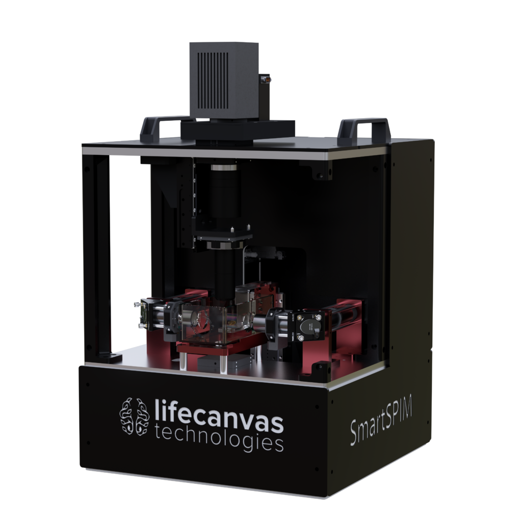
The SmartSPIM light sheet microscope provides unparalleled resolution and acquisition speed throughout intact tissue volumes, while its modular design allows you to upgrade and customize individual components.
Key highlights and features include:
The MegaSPIM microscope is our newest light sheet imaging system. Utilizing the same powerful axial sweeping technology as SmartSPIM, MegaSPIM is optimized for imaging not only laterally large samples, but thin slices and arrays of small tissues as well. Unique highlights and features include:

Advanced bioimaging instruments, such as our SmartSPIM and MegaSPIM microscopes, are capable of generating enormous amounts of invaluable 3D data. Analysis and processing of these data is simplified by leveraging high-performance storage technologies.
LifeCanvas developed MOSAIC, a data storage solution to seamlessly integrate into your workflow, providing large storage capacity, and effortless management.
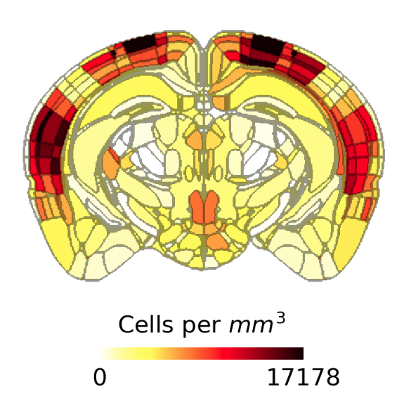
Quantify your results using our SmartSPIM-optimized workstation. Perform cell detection across the entire sample using machine learning pipelines customized and optimized for 3D fluorescent microscopy images. Generate detailed analysis outputs: visualize your effects with our publication-quality heatmaps, and statistically verify your experimental questions with per-brain region cell counts and densities for each sample.