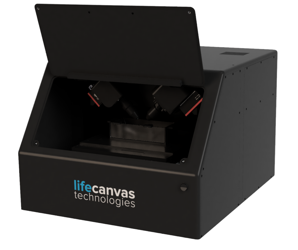
High-resolution light sheet microscopy for a wide range of applications
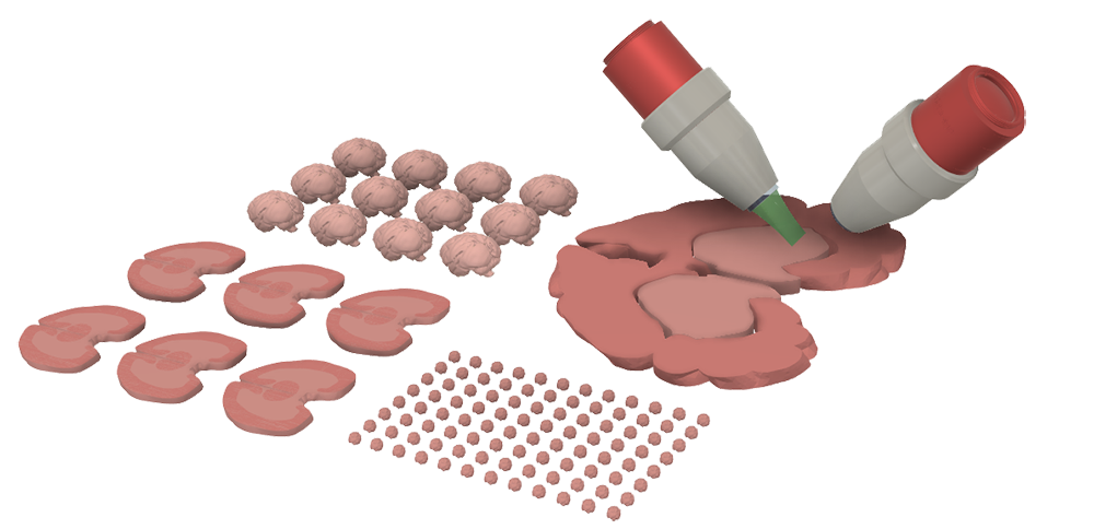
MegaSPIM is a highly flexible imaging system designed to rapidly acquire high-resolution data from a variety of tissue types, including: thin sections, cleared thick sections of human or nonhuman primate organs, and arrays of smaller cleared samples such as whole rodent organs or organoids. We offer custom mounting solutions for all of these tissues and more.
After image acquisition, our streamlined post-processing pipeline facilitates destriping, deskewing, and stitching large data sets. Easily create complete 3D image sets that are ready for analysis.
High-throughput volumetric imaging,
designed for speed and versatility
Easily scan through your tissues and transform raw images into complete 3D stacks ready for viewing and analysis. MegaSPIM allows you to efficiently generate high-quality data from multiple or larger samples. 1mm-thick human brain section (see below), can be completed in just 8 hours with the 1.8X objective and a single camera.
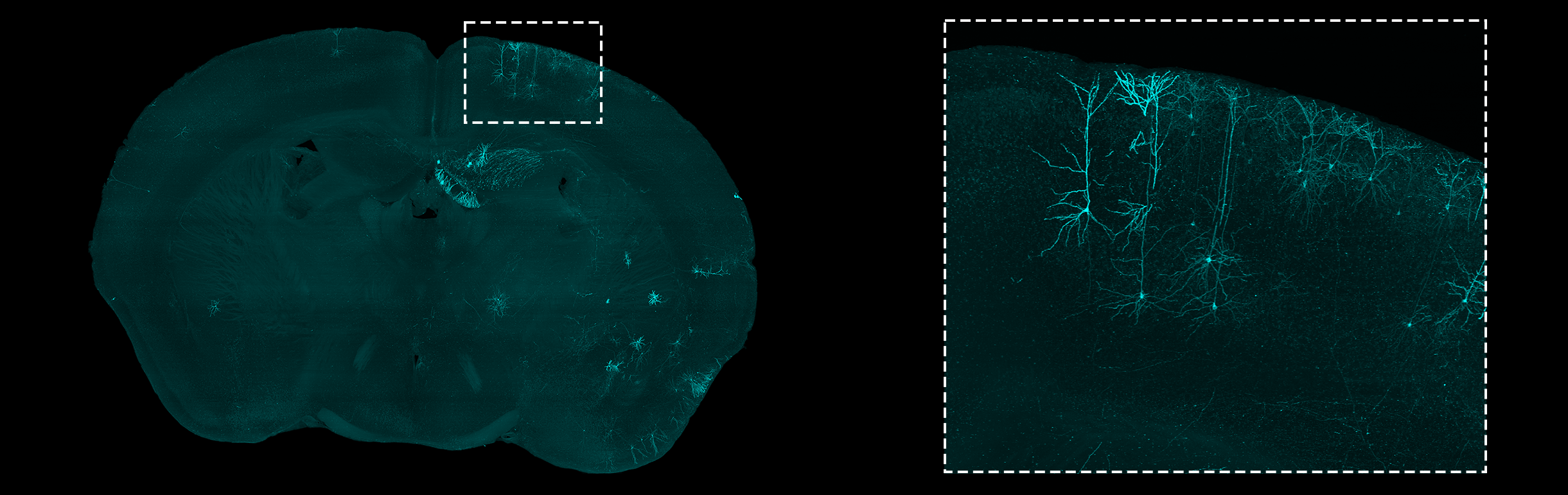
Detailed 3D data from human-scale samples to thin sections
TIGRE-MORF mouse brain, expressed via AAVI-Cre, imaged with MegaSPIM at 9X magnification. Shown: 200um Maximum Intensity Projection from a 1mm thick coronal section. Sample courtesy of Hongwei Dong Lab at UCLA.
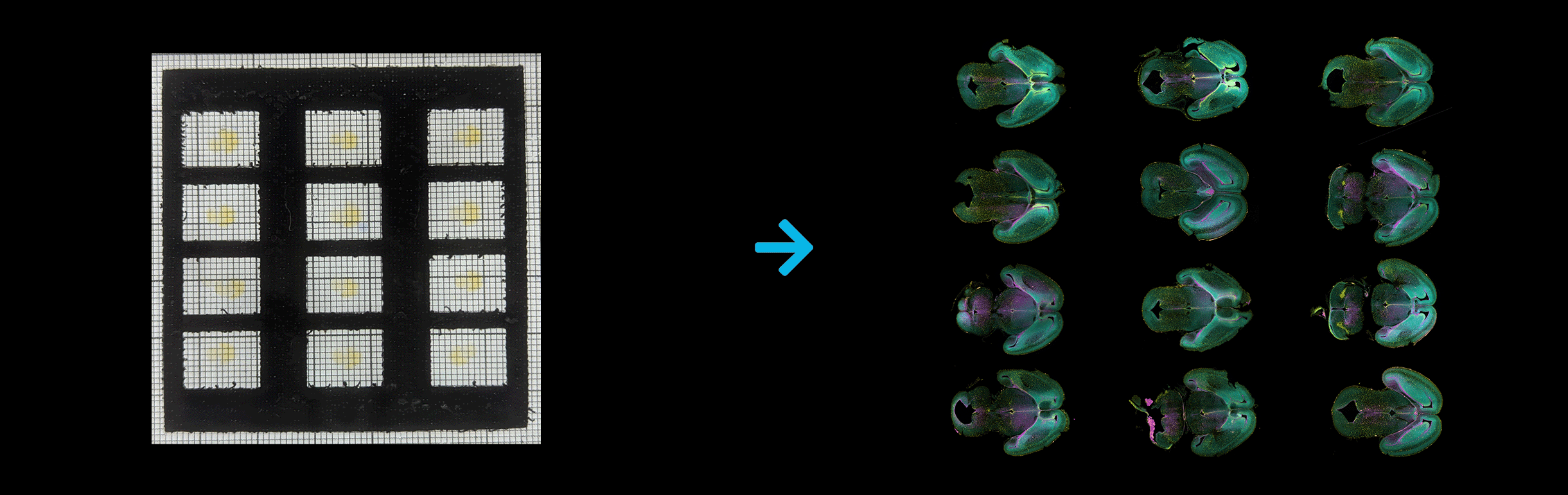
E14.5 mouse embryo brains labeled for cell nuclei via YoPro1 (cyan), Olig2 (yellow), and C-Fos (magenta). Courtesy of Dr. Claire-Marie Vacher, Columbia University. Imaged on MegaSPIM at 3.6X magnification, 30 minutes per sample (3 channels).
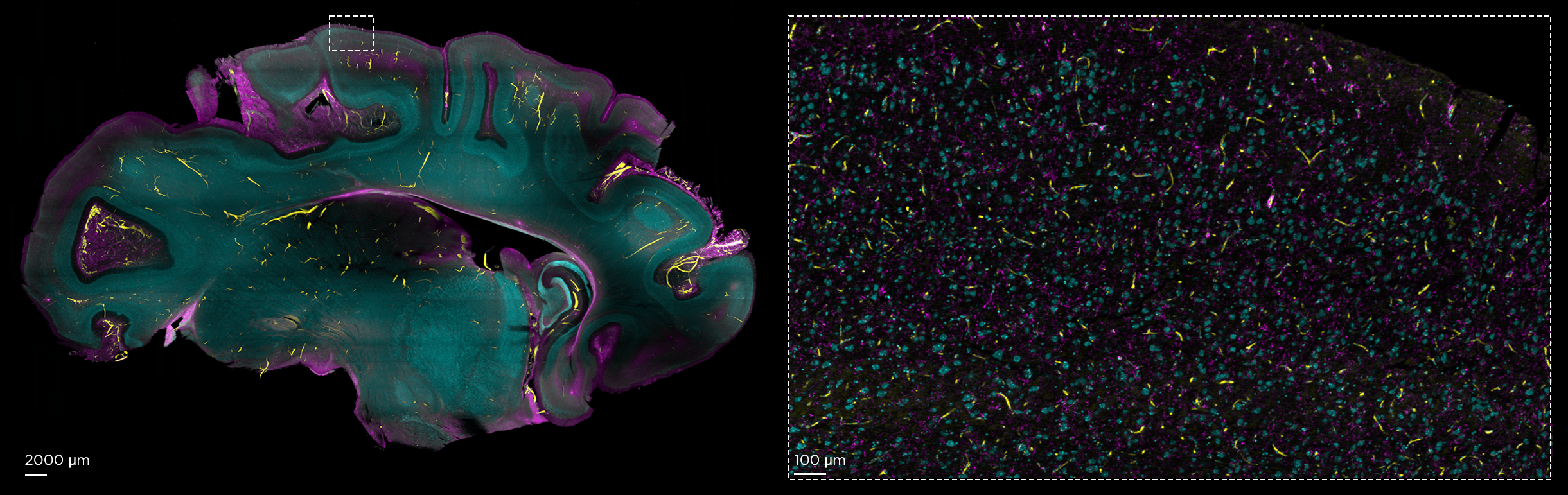
Syto16 (cyan), CD31 (yellow), + Iba-1 (magenta) in 2mm-thick section of pig brain, sliced with Megatome and imaged with MegaSPIM at 1.8X (left) and 15X (right) magnifications.
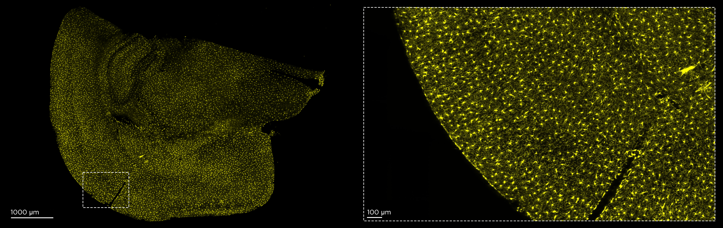
Iba-1 in 100µm-thick uncleared section of mouse brain, sliced with Megatome and imaged with MegaSPIM at 15X magnification.
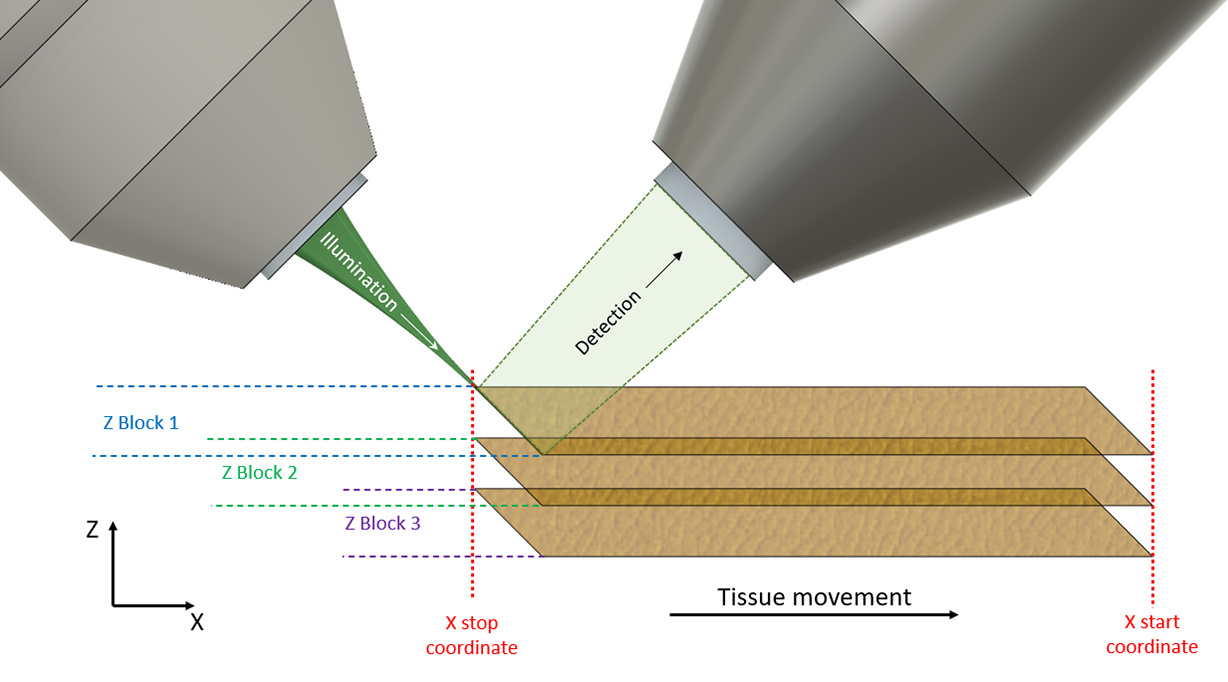
Light sheet illumination and detection
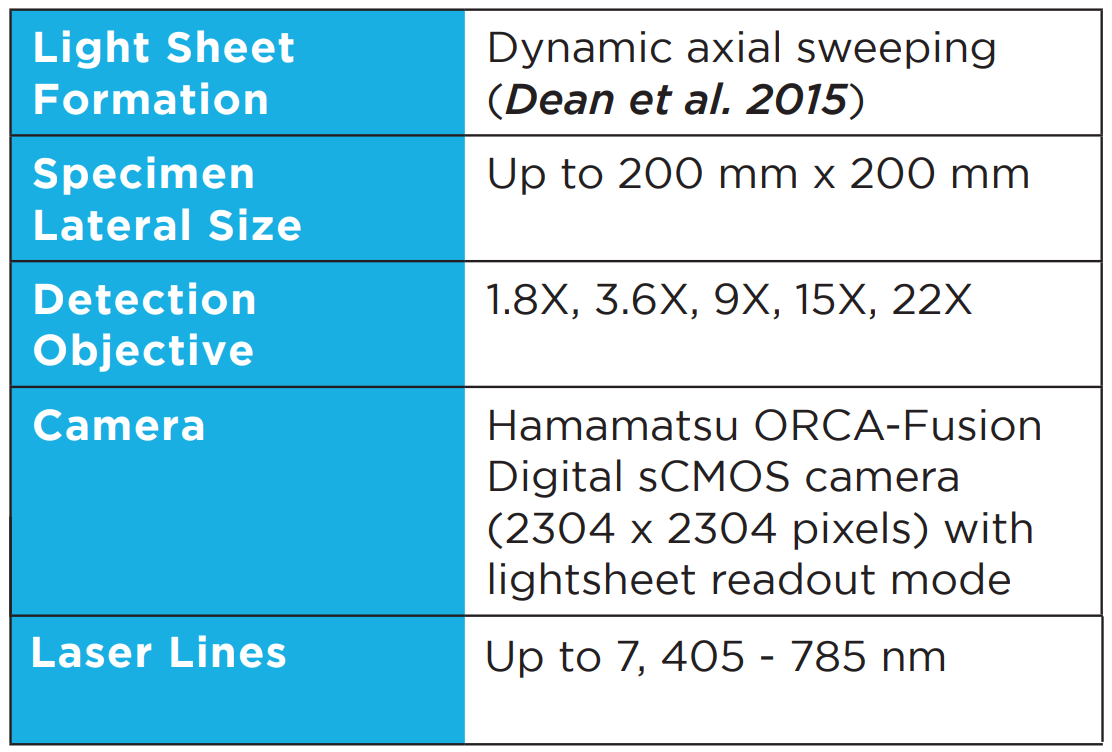
MegaSPIM uses patented axial sweeping technology to capture images with uniform axial resolution across the entire field of view without deconvolution. Long working-distance objectives positioned at 45 degrees above the sample plane allow imaging of samples as laterally large as the travel range of the stage.
Our deskewing algorithm seamlessly translates these tilted image stacks into standard XY planes, ready to be stitched into complete XYZ stacks for visualization and analysis.