
Illuminate the possibilities of biomedical research with optimized, integrated workflows for whole-tissue histology, imaging, and analysis. Whether you adopt our innovative tools in your own lab, or hand off your samples to our exceptional CRO technicians, new discoveries await.
Pictured above: MC3R-expressing neurons in hippocampus of intact mouse brain, imaged with SmartSPIM. Image courtesy of Dr. Michelle Bedenbaugh, Vanderbilt University.
Viral injection tracing methods, such as those utilizing rabies and AAV vectors, can be used to visualize neuronal connections throughout the brain and other innervated organs. Our tools offer a detailed 3D view of these connections at multiple scales, with no need for immunolabeling of endogenous fluorophores.
Recently published: BNST PKCδ neurons are activated by specific aversive conditions to promote anxiety-like behavior (Williford et al. 2023)
Our tools can provide unbiased insights into the pharmacodynamics of different therapeutic delivery methods, including AAV vectors, lipid nanoparticles (LNPs), small molecules, and mAbs. Researchers can obtain high-resolution biodistribution data at the cellular level with detailed spatial coverage, and the ability to multiplex markers for different target cell types.
Our technologies offer researchers an unbiased look at beta-amyloid plaque distribution throughout whole brain volumes, as well as interactions between plaques and other cell types such as microglia. Detailed 3D mapping of plaques with other neuronal cells reveals valuable information about molecular disease phenotypes, such as disease progression and immune response.
Recently published: Loss of insulin signaling in astrocytes exacerbates Alzheimer-like phenotypes in a 5xFAD mouse model (Chen et al. 2023)
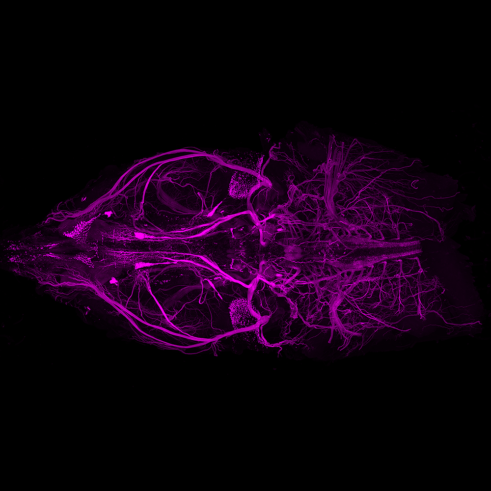
Understanding the intricacies of vascular networks is key to painting a more complete picture of critical processes in development, disease, and injury. Our 3D vasculature mapping techniques preserve connectivity of blood vessels, resolve fine structures, and provide unmatched spatial data.
Recently published: Aging drives cerebrovascular network remodeling and functional changes in the mouse brain (Bennett et al. 2023)
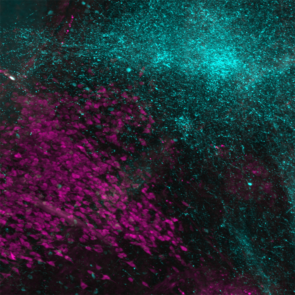
Metabolic processes are driven and regulated by a complex network of interactions between body systems. High quality spatial data can reveal crucial, often underappreciated connections between the nervous system and adipose tissue, as well as other systems that impact metabolism and related disorders such as obesity and diabetes.
Recently published: The role of somatosensory innervation of adipose tissues (Wang et al. 2023)
Tumors are highly heterogenous tissues and are thus not well-sampled by 2D, sectioning-based methods. Our tools allow researchers to obtain 3D data from intact tumors, providing a more accurate, spatially unbiased approach to studying diseased tissues.
Traditional 2D sectioning approaches to studying brain injury can be tedious and destructive, as well as limited in their representation of contiguous structures. Accurate 3D visualization and molecular characterization of brains exhibiting traumatic brain injury (TBI) unlocks a whole new dimension of insights for disease models as well as clinical samples.
Recently published: Leveraging the Power of 3D Brain-Wide Imaging and Mapping Tools for Brain Injury Research in Murine Models (Anwer et al. 2023)
A well-recognized hallmark of Parkinson’s disease is the presence of Lewy bodies, which are composed primarily of the aggregation-prone neuronal protein α-Synuclein. 3D mapping of α-Synuclein in diseased tissues gives scientists an unparalleled opportunity to examine brain-wide changes in and characteristics of Parkinson’s disease.
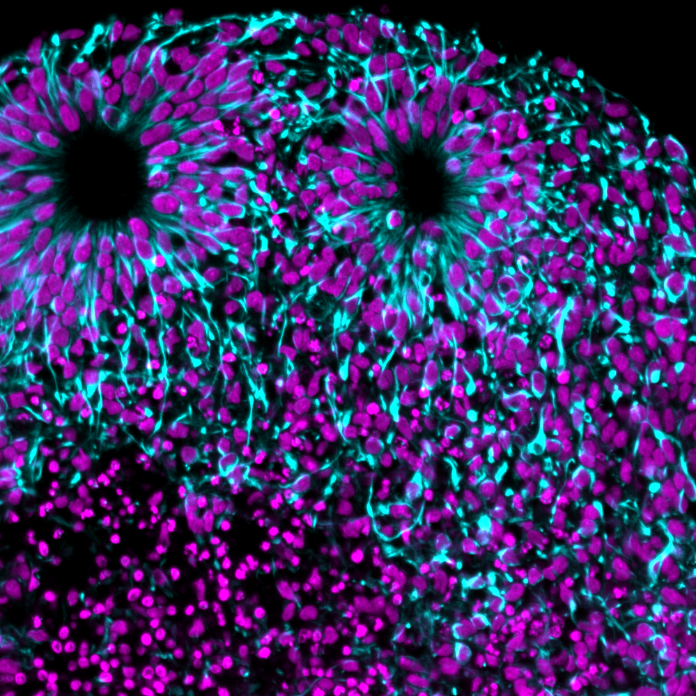
Organoid models of mammalian tissues allow researchers to closely study the development of highly organized systems from stem cells, and how those processes go awry in disease. LifeCanvas tools enable high-throughput processing and imaging of organoid arrays, accelerating the process of characterizing and garnering insights from samples.
Recently published: Dephosphorylation of 4EBP1/2 Induces Prenatal Neural Stem Cell Quiescence (Geben et al. 2023)
We are always seeking opportunities to expand and adapt our pipeline, and empower more researchers across the biomedical sciences. Get in touch with us about your ideas – let’s work together to elevate human health.