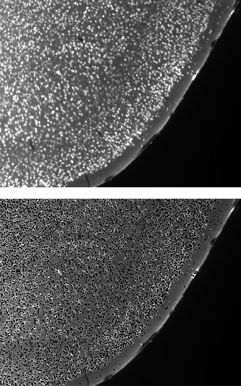
We use deep learning models to accurately detect and quantify diverse cell types from light sheet data. For neuroscience studies, automated mouse and rat brain atlas registration gives you access to in-depth regional information.
Our analysis methods are optimized for LifeCanvas-quality light sheet datasets, providing detailed and precise insights for many downstream applications. Leverage quantitative analyses from per-region cell counts and signal intensity to co-localization.
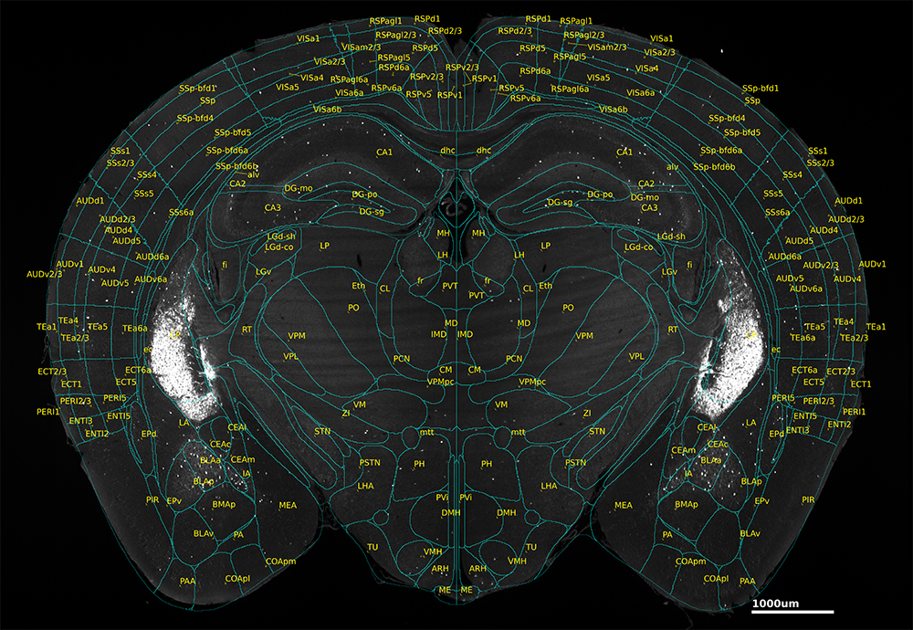
You don’t have to be a computer scientist or expert neuroanatomist to produce a well-aligned dataset. We provide automated alignment to mouse and rat brain atlases, enabling precise cell counts and fluorescence localization measurements.
Publication-ready results
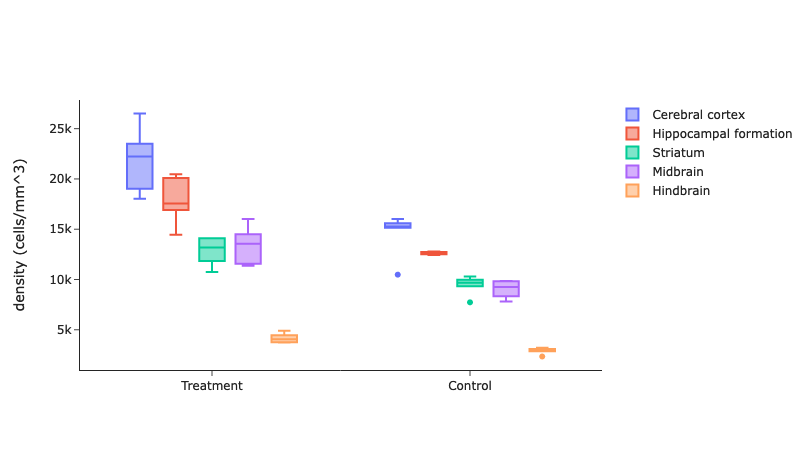
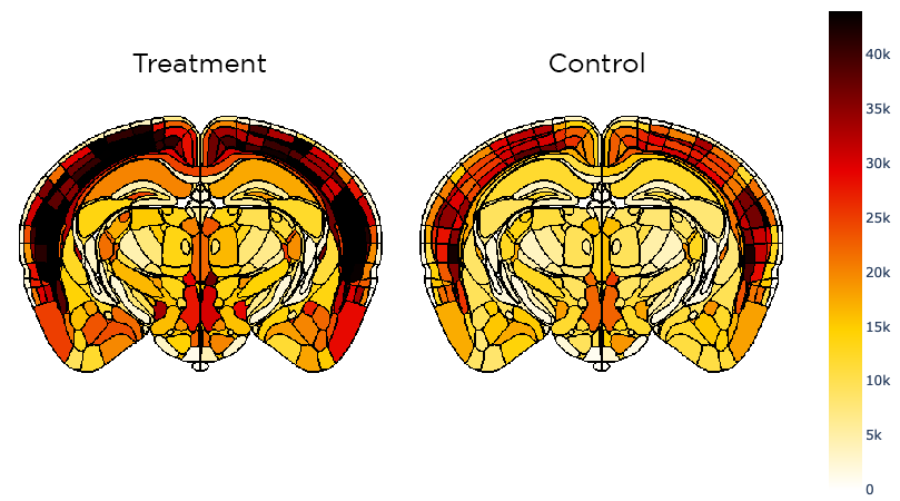
We can generate accurate cell counts and fluorescence intensity outputs for highly granular brain regions, enabling multi-sample statistical significance testing. Download heat maps directly from our DataCanvas portal, ready to be embedded into your next grant submission, presentation, or publication.
Adaptable cell detection models
Machine learning algorithms provide reliable cell detection for a variety of applications, including:
Neural activity: Detect and map c-FOS+ cells in whole brains for insight into brain regional activity levels unaffected by selection bias.
Endogenous fluorescence: Identify and quantify fluorescently-tagged cells throughout whole tissue volumes.
Neurological disease markers: Assess disease progression and expression, as well as neuroinflammatory response, with models for astrocytes (GFAP) and microglia (IBA1), and segmentation analyses for β-amyloid deposits (senile plaques) and α-synuclein aggregates (Lewy Bodies).
Other cell-type markers: Identify and quantify neurons immunolabeled for NeuN, TH, PV, and more.