3D Phenotyping with the SmartSPIM Light Sheet Microscope
After tissue clearing and (optional) labeling, intact tissue needs to be index-matched with EasyIndex refractive index-matching solution. Afterwards, tissues will appear fully transparent and ready for imaging using the SmartSPIM light sheet microscope. To complete your workflow, LifeCanvas Technologies offers SmartAnalytics – a powerful machine learning analysis tool, specially designed for large 3D datasets.
This blog will provide key insights on the last steps to complete your tissue processing journey: refractive index-matching with EasyIndex, volumetric imaging with SmartSPIM, and image analysis with SmartAnalytics.
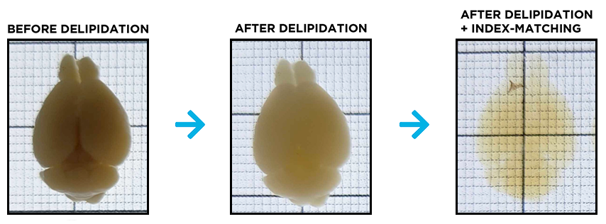
Refractive Index-matching with EasyIndex
To reach complete tissue transparency, refractive indices need to be matched between the tissue and the immersion medium used for imaging. A few hours of incubation in EasyIndex refractive matching solution readily results in complete refractive index-matching. This renders the tissue transparent and reduces light scattering, allowing for high-resolution imaging throughout the entire tissue volume.
For subsequent imaging, it is crucial that samples are immobilized. Other imaging set-ups offer a holder with screws on the side or suggest gluing the sample to the holder. While these may help with stabilization, the screw and glue can interfere with imaging or cause subtle movement, leading to light refraction. LifeCanvas has designed a unique sample holder that enables embedding of specimens in a low melting point agarose gel composed of EasyIndex, securing the sample in the holder without the interference of additional screws or glue.
Volumetric imaging with the SmartSPIM light sheet microscope
Our light sheet microscope is an indispensable component of our intact tissue molecular phenotyping pipeline. In contrast to point scanning with confocal microscopy, light sheet microscopes focus excitation light on thin planes of illumination across a wide field of view perpendicular to the axis of the imaging objective. This arrangement substantially improves the resolution and speed of image acquisition from large, cleared volumes. Imaging with a light sheet microscope also minimizes out-of-focus exposure, while maximizing axial resolution and acquisition speed.
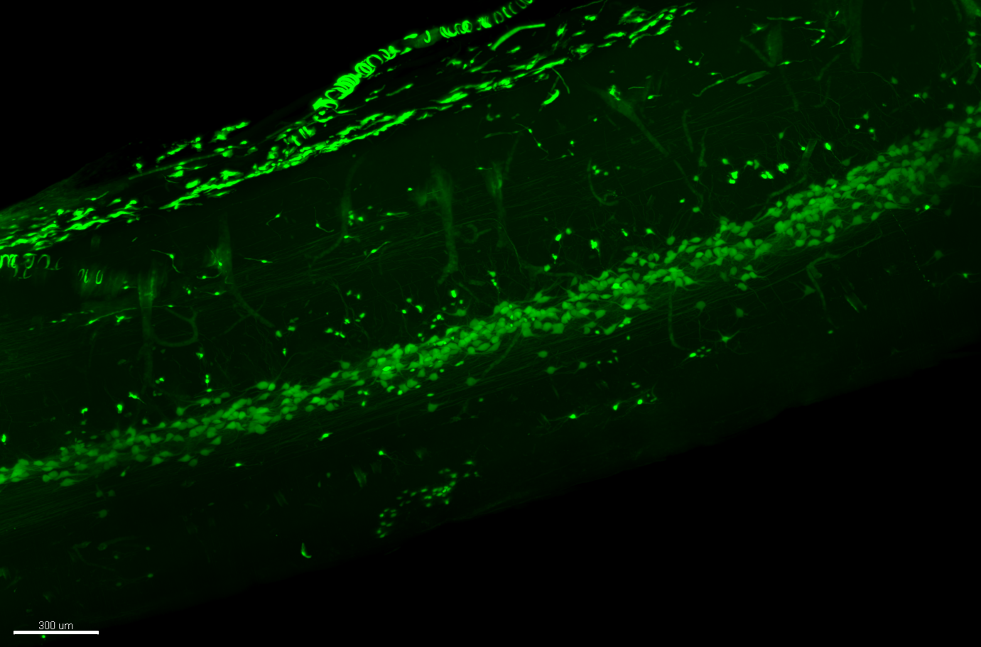
Mouse spinal cord expressing tdTomato. Sample courtesy of Lai Lab, UT Southwestern.
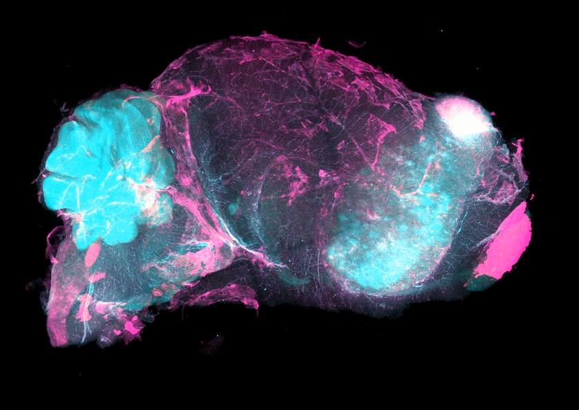
Nuclear stain (SYTO16, in cyan) and vasculature stain (DyLight 649-conjugated tomato lectin, in pink). Tissue provided by Translational Bioimaging Group at the Barrow Neurological Institute.
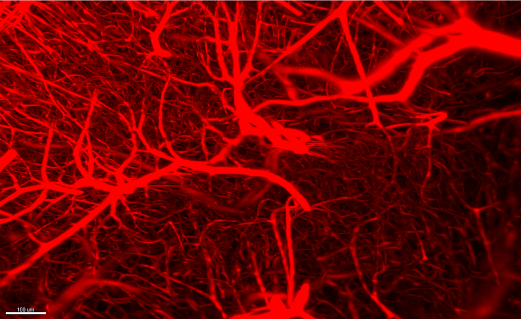
Tissue stained with lectin dye, which labels blood vessels with high specificity and SNR.
15X, 1.5 x 1.5 x 1.8 mm region acquired at 1 µm Z-step in <5 minutes.
LifeCanvas designed and built a dynamic, flexible light sheet microscope based on patented axially-swept light sheet technology (Dean et al. 2015). The SmartSPIM light sheet system is optimized for high resolution and high speed image acquisition from large cleared tissues. SmartSPIM’s imaging chamber and sample holders are customized and suitable for a wide range of tissues, from organoids to whole rodent brains, and even entire juvenile mice.
SmartSPIM’s axially-swept light sheet is synchronized to the rolling shutter of the sCMOS camera, resulting in confocal-like detection with uniform and improved single-cell resolution. Fast acquisition enables imaging of large tissues in minutes to hours – considerably faster than the potentially days-long acquisition rates of other light sheet microscopes.
SmartSPIM’s acquisition software is also user-friendly and designed for efficient and reliable data collection with minimal training required.
Advanced 3D image analysis with SmartAnalytics
To complete LifeCanvas’ end-to-end tissue processing pipeline, the SmartAnalytics workstation offers Allen Brain Atlas alignment, cell detection, and fluorescence intensity quantification. This advanced machine-learning analysis tool has been designed specifically for tackling large (i.e. terabyte-sized) 3D image datasets.
SmartAnalytics software enables easy and accurate alignment of murine brain images to the Allen Brain Atlas along with detection, mapping, and quantification of many key neuroscience markers, such as GFAP (astrocytes), IBA1 (microglia), and neuronal cell markers including NeuN, tyrosine hydroxylase, parvalbumin, and neuropeptide Y.
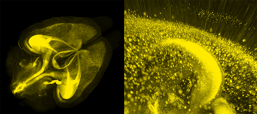
Thy1-driven YFP expression in whole mouse brain. Sample courtesy of G. Allan Johnson, Duke Center for In Vivo Microscopy.
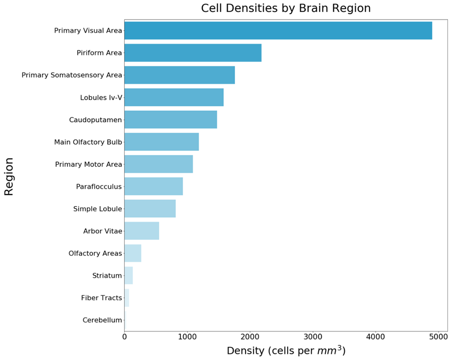
SmartAnalytics also allows the detection and mapping of neural functional activity. After clearing, immunolabeling, and imaging of c-FOS+ cells in whole mouse brains, LifeCanvas provides customers with quantification of neural activityvia cell counts, densities, and heatmaps of c-FOS+ cells throughout the entire brain.
Hippocampus and cortex in sagittal plane of mouse brain hemisphere stained with anti-c-FOS.
Histogram of c-FOS+ cell density by region in mouse brain hemisphere.
Heatmap of c-FOS+ cell density by region in mouse brain hemisphere.
New to 3D Whole Organ Mapping?
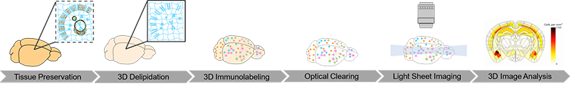
LifeCanvas Technologies offers exceptional-quality Contract Research (CRO) service for tissue preservation, clearing, immunolabeling, refractive index-matching, light sheet imaging, and image analysis, all tailored to your scientific needs.
Multiple research groups have published data using the CRO services, including:
The LifeCanvas scientific team has extensive experience with multiple tissue types including mouse intestine, kidney, brain, spleen, liver, heart, embryos, spinal cord, and colon; rat brains and lungs; human skin and lungs; as well as organoids and tumor samples.
Versatile immunolabeling is also offered for common targets for neural activity (mapping and quantifying c-FOS+ cells), Alzheimer’s disease (β-amyloid deposition), Parkinson’s disease (α-synuclein Lewy Bodies), and neural inflammation (IBA1 for microglia and GFAP for astrocytes).
LifeCanvas prepares your samples for volumetric imaging with SmartSPIM, followed by holistic analyses of the tissue with SmartAnalytics. High quality, publication-ready results are then delivered to you for your next grant submission, presentation, or publication!
Conclusion
LifeCanvas Technologies’ products enable fast advanced tissue clearing, labeling, imaging and analyses, resulting in high quality, reliable data. The team’s scientific experts continually go above and beyond to offer exceptional customer support and service throughout your samples’ entire journey through our pipeline. Contact us to discover how LifeCanvas can support your research goals!