Published by Park et al. in Nature Biotechnology 2018, our SHIELD reagent (AKA GE38) safeguards endogenous fluorescence, protein antigenicity, nucleic acids, and overall tissue architecture against physical and chemical stressors. The SHIELD tissue preservation method also protects tissue during fluorescent protein imaging and antibody labeling across multiple rounds.
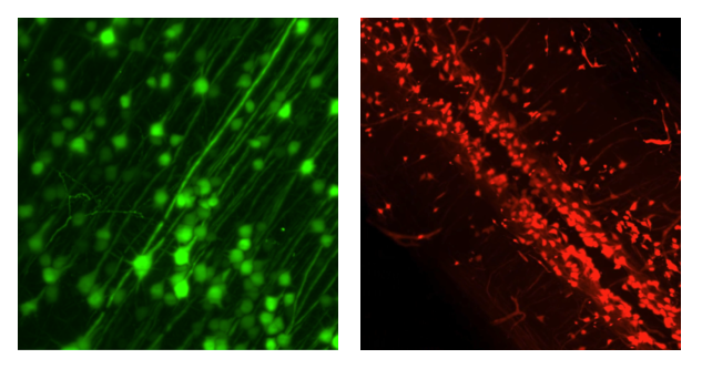
Left: GFP+ motor cortical neurons, sample courtesy of Dr. Byungkook Lim, UCSD. Right: RFP+ spinal cord cells, sample courtesy of Dr. Helen Lai, UT Southwestern. Both imaged with SmartSPIM.
Quick & easy tissue preservation protocol
Our streamlined protocol takes just 4-6 days, without the variability of tissue embedding via hydrogel or paraffin. Reagents are non-toxic and can be easily disposed of after use.
Mohammed A. Khallaf, Daniel W. Hart, Wenhan Luo, Firdevs Murad, Felipe Cybis Pereira, Daniel Mendez-Aranda, Nicole Hagenah, Alice Rossi, Valérie Bégay, Jan Okrouhlík, Dietmar Krautwurst, Mungo Kisinza Ngalameno, Andre Ganswindt, Alison J. Barker, Radim Šumbera, Markus Knaden, Sophie Pezet, Andrew Woehler, Bill S. Hansson, Nigel C. Bennett & Gary R. Lewin
Yehui Sun, Yu-Chung Pien, Yufen Xiao, Wei Tan, Efrain Sanchez-Ortiz, Mateusz Z. Durbacz, Zexiang Chen, John R. McAnally, Yu Zhang, Hui Li, Andreas C. Chai, Francesco Chemello, Fangyu Zhang, Sang M. Lee, Priyanka Patel, Denise M. Ramirez, Sumanta Chatterjee, Ning Liu, Eric N. Olson, Daniel J. Siegwart