Recent advances in the tissue clearing field have unlocked the visualization of biology throughout whole organs. Tissue processing methods that render samples optically transparent can be combined with methods that enable the tissue-wide penetration of molecular probes such as antibodies. However, many of these techniques – which can include dehydration, delipidation, high temperature exposure, and protease treatment – are harsh and can lead to the significant sample degradation and loss of biomolecular information.
To protect tissue samples against damaging physical and chemical treatments, LifeCanvas scientists use a novel tissue transformation system called SHIELD (Stabilization to Harsh Conditions via Intramolecular Epoxide Linkages to Prevent Degradation).
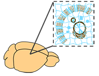
This versatile method employs a unique polyepoxide that cross-links molecules within biological specimens, thus creating a reinforced tissue-gel (Fig. 1).
As a result, biomolecules are secured at their physiological locations and tissue mechanical stability is greatly enhanced. The SHIELD polyepoxide molecule owes its effectiveness as a cross-linking agent to its multiple functional groups and flexible polyglycerol backbone. These properties enable binding not only to multiple sites within the same molecule but also the formation of cross-links with neighboring biomolecules. The polyepoxide’s flexible backbone can additionally accommodate thermal fluctuations and preserves protein conformation. As such, SHIELD-preserved tissues can effectively maintain their native tissue architecture and biomolecular integrity under the wide range of harsh conditions that is required for tissue clearing and labeling techniques.
SHIELD represents a real improvement in sample preservation for tissue clearing. Although exposure to organic solvents and detergents can lead to a dramatic loss in fluorescent protein signal in standard PFA-fixed tissues, SHIELD preservation can largely retain fluorescence under these same conditions. SHIELD-preserved tissue is optimized for 3D immunolabeling because antigenicity is well preserved and compares favorably with other approaches. Importantly, the SHIELD epoxide can form covalent bonds with nucleic acids thus enabling in situ hybridization approaches. Autofluorescence is kept at a minimum in SHIELD-ed tissues. Moreover, various tissue clearing protocols can lead to sample deterioration and shrinkage whereas SHIELD can best maintain the size and shape of the original sample. This faithful preservation of tissue morphology is critical for accurate registration and analysis of datasets obtained from whole brain, volumetric imaging.
SHIELD also has many other practical benefits. SHIELD-preserved tissues can be rapidly cleared and immunolabeled using stochastic electrotransport (SE), a rotational electric field which accelerates the dispersion of electromobile molecules. Clearing and labeling protocols which once took weeks only require a few days with SE-powered technology such as SmartBatch+, SmartClear II Pro, and SmartLabel. SHIELD is also easy to use, can completely preserve rodent organs (e.g. whole mouse brains) within a matter of days, and can be readily scaled up for labs that demand higher throughput. Because of greatly improved tissue stability, SHIELD-preserved tissues can be repeatedly interrogated with multiple rounds of immunolabeling. SHIELD has also been shown to be compatible with intact biopsy samples. Because of its many advantages, LifeCanvas Technologies exclusively uses SHIELD for the essential sample preservation step of our end-to-end 3D histology workflow.