3D AAV Biodistribution Mapping

3D spatial profiling for AAV gene delivery

AAV-mediated gene delivery is a promising therapeutic approach for many diseases, from cancer to rare genetic disorders. With our advanced tools for 3D histology, imaging, and analysis, LifeCanvas generates unbiased biodistribution data to evaluate AAV delivery throughout intact tissues. Our technologies are applicable to a variety of tissue types, including: whole organs such as brain, heart, and lung, unique samples like biopsies, tumors, and organoids, and even whole juvenile mice.

Pictured above: Endogenous tdTomato in whole adult mouse brain, imaged with SmartSPIM at 3.6X magnification. Sample courtesy of Dr. Shenfeng Qiu, University of Arizona College of Medicine.

Icons highlighting different organs: brain, ear, liver, GI tract, eye, lungs, heart, spinal cord, whole mouse
Figure showing, left to right: GFP+ motor cortical neurons in whole mouse brain, tdTomato+ cells in intact mouse spinal cord, Thy1-driven YFP expression in whole mouse brain
Left to Right: GFP in mouse cortex, tdTomato in mouse spinal cord, YFP in mouse brain.

Visualize fluorescent protein distribution throughout tissues

Superior signal preservation enables sensitive detection of fluorescent protein expression (e.g. GFP, tdTomato, YFP) driven by virally-delivered genes. Our proprietary SHIELD method protects biomolecules during downstream steps, and our Clear+ tissue clearing technique maintains fluorescent signal, allowing for high-resolution light sheet imaging with SmartSPIM or MegaSPIM.

Evaluate targeted gene delivery with cell-type co-localization data

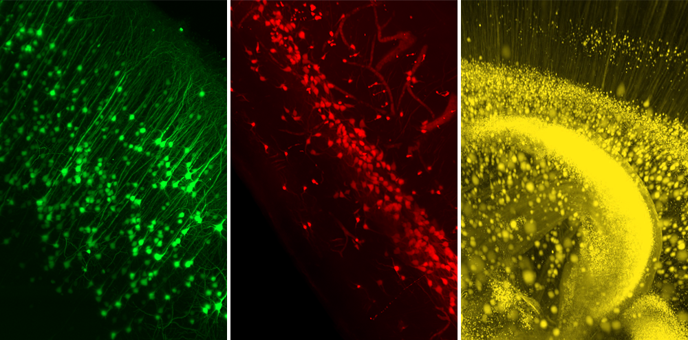
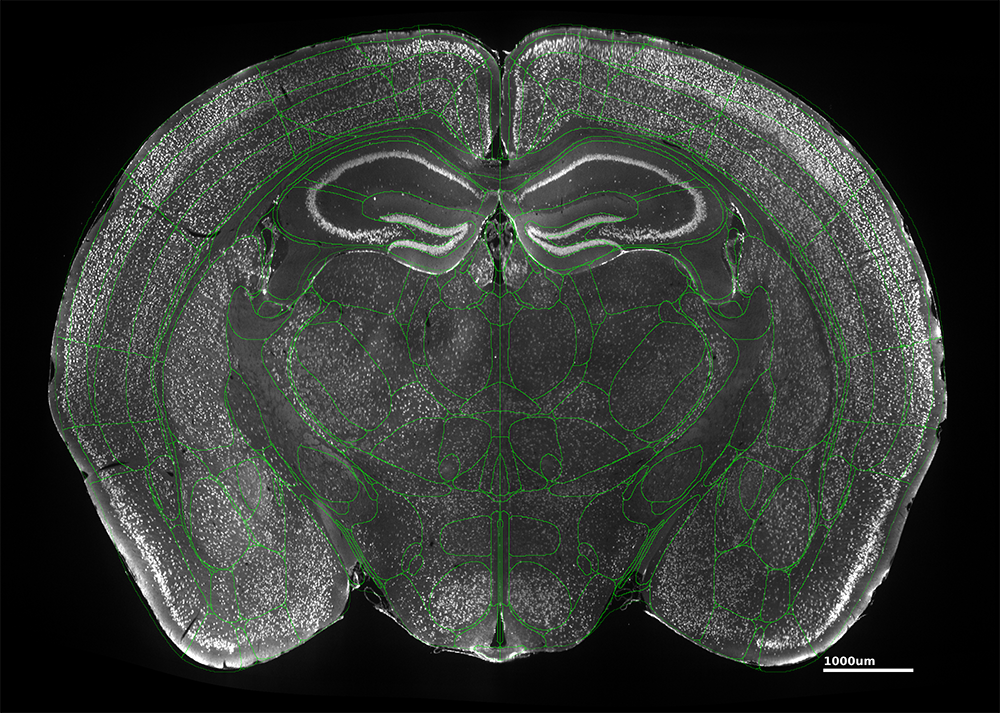
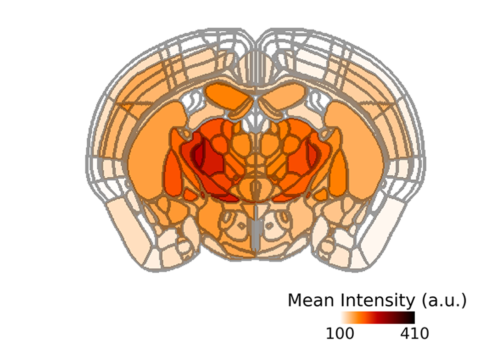
3D bioimaging can determine cell type specificity of capsids by co-localizing fluorescent protein signals with cell-type labeling. This capability is enabled by eFLASH technology which ensures robust, uniform labeling of neurons, astrocytes, and microglia using cell-type markers (e.g. NeuN, GFAP, IBA-1) throughout whole tissues. 

Illustration of neuron, microglia, and astrocyte

Comprehensive analysis outputs

Our SmartAnalytics workstation generates robust outputs from large 3D image datasets, including cell counts, fluorescence intensity mapping, and co-localization. AI-powered models allow reliable detection and quantification of FP-tagged cells as well as different cell types, while registration to the Allen Brain Atlas or Waxholm Space Atlas ensures precise analysis of whole-brain data.

To streamline data delivery, our interactive web platform allows you to easily visualize outputs with customizable heat maps and bar plots.

Speak to an expert about your application