Precision sectioning of human and non-human primate brains with Megatome Premium
Introduction
Neuroscience research based on the human and non-human primate (NHP) brains has uncovered significant knowledge about the brain function, disorders and treatment. However, collecting cellular/subcellular resolution images of the human and NHP brains is extremely labor-intensive and time-consuming using conventional immunohistochemistry (IHC) methods, which commonly involves specimen blocking, freezing, thin-sectioning, as well as considerable amount of tissue loss and damage. It’s almost unthinkable to map the whole human or NHP brains based on these processes.
As a pioneer in 3D histology, LifeCanvas Technologies has stepped forward with new tissue processing products and high-throughput solutions for 3D phenotyping of human and NHP brains. Here, we demonstrate how the Megatome Premium can easily facilitate this process through whole-mount and non-destructive tissue sectioning.
Whole-mount, high-throughput sectioning
Human and NHP brains are significantly larger and anatomically more complex than rodent brains. Due to limitations of the majority of tissue sectioning devices and their complexity of operation, researchers are only able to process small blocks of tissues picked from different parts of the brain, leaving important information such as spatial correlations and connectivity networks behind.
Megatome Premium is the only tissue sectioning device that allows for whole-mount, high-throughput sectioning of the human and NHP brains. It saves researchers a huge amount of time and produces unprecedented high quality tissue sections.
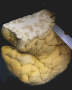
Below are examples of using the Megatome Premium to generate tissue sections from the whole human cerebral hemisphere, human cerebellar hemisphere, and the whole marmoset brain.
Figure 1. Human cerebral hemisphere embedded in gel (left); and being sectioned by Megatome (right).
Figure 2. Human cerebral hemisphere coronal sections in 5 mm thickness.
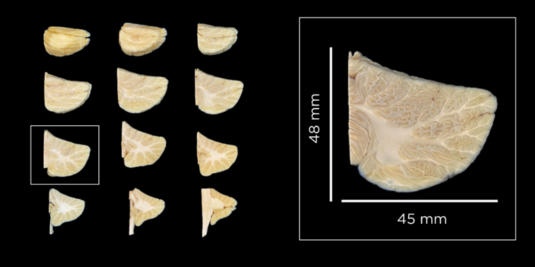
Figure 3. Human right cerebellum and brainstem coronal sections in 5 mm thickness.
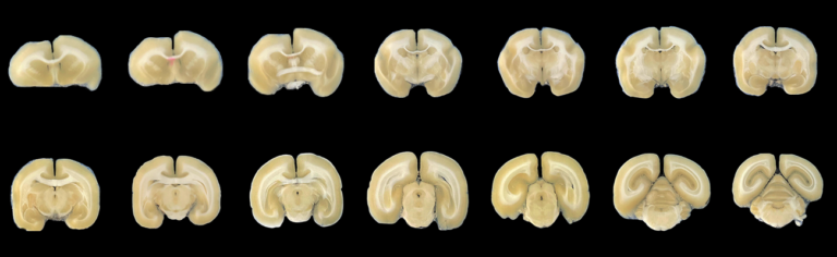
Figure 4. Whole marmoset brain coronal sections in 1 mm thickness.
Non-destructive, thin sectioning
Precision sectioning is another important feature of the Megatome Premium, which allows users to produce intact, thin and large tissue sections. Small samples such as the rodent brain tend to have more uniform mechanical textures, making it easier to produce thin sections (< 100 µm) for traditional IHC processes (versus tissue clearing processes that can label much thicker samples). However, the mechanical inhomogeneity and significantly large dimensions make it much more challenging to obtain high quality, thin sections.
Thanks to the high blade vibration frequency, amplitude, minimized blade deflection, and high accuracy sample translation movement, the Megatome Premium is able to section tissues exceptionally smoothly and thinly causing no tissue damage or loss.
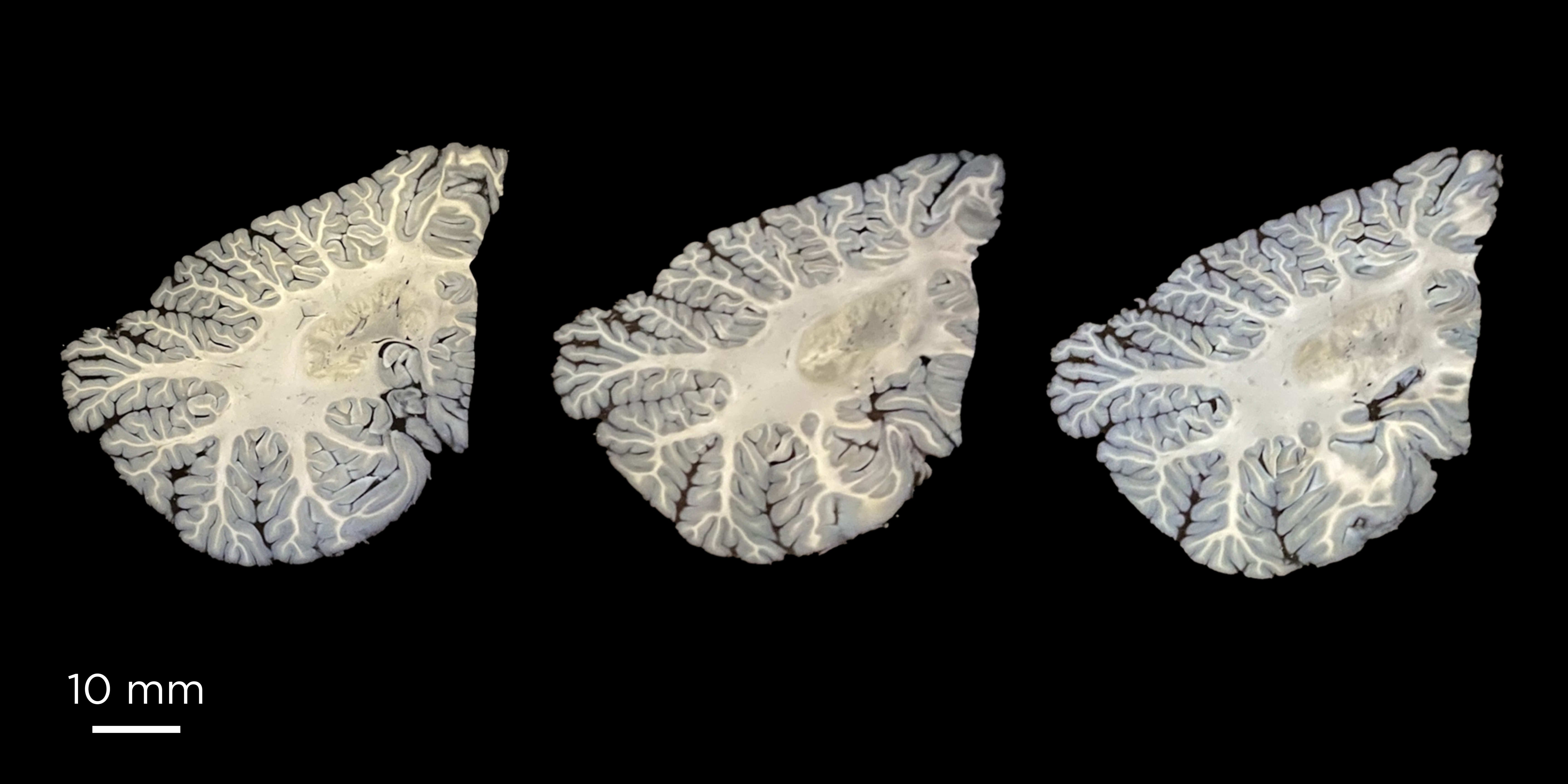
Examples below demonstrate non-destructive thin sections of large and delicate human samples produced by the Megatome Premium. Fine structures such as the tightly folded cerebellar cortex and the inner structure of the medulla oblongata are clearly reviewed from the sections.
Figure 5. Human cerebellum coronal sections in 100 μm thickness.
Figure 6. Human pons, medulla oblongata and spinal cord coronal sections in 50 μm thickness.
Megatome & tissue clearing, labeling and MegaSPIM imaging:
Megatome Premium together with LifeCanvas tissue preservation and clearing reagents and MegaSPIM imaging system represents a complete solution for human and NHP tissue high-throughput multi-scale imaging and phenotyping.
Example below demonstrates the above solution with a marmoset brain injected with CTB-Alexa Fluor561 and CTB-Alexa Fluor 647. The whole marmoset brain was first sectioned with Megatome Premium into intact 1 mm thick sections. The sections were then processed with SHIELD, clearing, index matching, and finally imaged with MegaSPIM (see images below).
Original Marmoset Brain
After SHIELD
After Clearing
Bottom images, from left to right: 1 mm marmoset slab with CTB injections targeting dorsal raphe and hippocampus in 561 nm (magenta) and 647 nm (cyan); cortical layers; aqueduct and dorsal raphe targeted with CTB injection in 561 nm (magenta) and 647 nm (cyan) channels. Samples courtesy of Denis Matrov and the Yogita Chudasam Lab at National Institutes of Health.
Summary
Megatome Premium is the most versatile tool for generating sections from human, NHP, and small animal samples with extremely high throughput and high quality. It not only helps researchers save time and effort in preparing samples, but also opens new opportunities to study the brain in more dimensions, revealing more spatial, molecular, morphological, and connectivity information of the brain.