Immunohistochemistry (IHC) and 3D histology are different approaches to detecting specific proteins in biological tissue.

Features and highlights:
Video shows how SeTau-647 maintains photostability over time compared to a conventional 647 fluorophore. A whole mouse brain was labeled with anti-NeuN primary antibody. After hemisecting the brain, the hemisphere on the left was stained with a conventional 647 fluorophore secondary antibody, and the hemisphere on the right with SeTau-647. Hemispheres were glued together and imaged on SmartSPIM at 1.6X with 100% laser power.
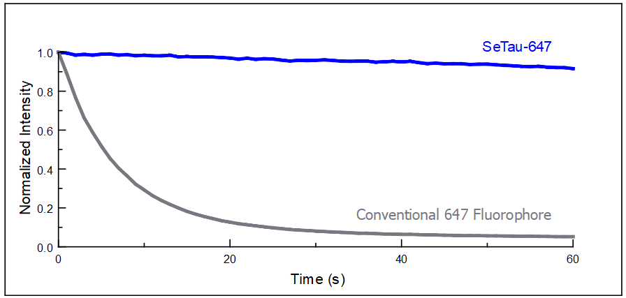
Photobleaching occurs when a fluorophore loses its fluorescence due to light-induced damage. As different fluorophores have varied susceptibility to this phenomenon, we measure fluorescence intensity of different fluorophores over time to determine photostability.
LifeCanvas developed SeTau-647 Secondary Antibody which as observed in the graph, shows higher fluorescence intensity sustainability over time compared to conventional 647 fluorophores. This establishes SeTau-647 as the premier choice, showcasing superior photostability over conventional alternatives.
Minimizing photobleaching is crucial for light sheet imaging and other applications requiring fluorescence stability, such as 2P, expansion tissues, super-resolution, and live cell imaging.
Lindsay L. Ejoh, Raquel Adaia Sandoval Ortega, Jacqueline W. K. Wu, Alaa Elhiraika, Malaika Mahmood, Kyungdong Kim, Emmy Li, Oliver Joseph, Lisa M. Wooldridge, Jessica A. Wojick, Gregory J. Salimando, Gregory Corder
Zuri A Sullivan, Bertrand J. Wong, Vikrant Kapoor, Paula Kathleen Vincze, Harris S. Kaplan, Pia Giraudet, Brianna R. Watson, Adel Misherghi, Sebastian Lourido, Jeffrey R Moffitt, Catherine Dulac
Quan Jiang, Ling-Xiao Shao, Shenqin Yao, Neil K. Savalia, Amelia D. Gilbert, Pasha A. Davoudian, Jack D. Nothnagel, Guilian Tian5, Tin Shing Hung, Hei Ming Lai, Kevin T. Beier, Hongkui Zeng, Alex C. Kwan
Immunohistochemistry (IHC) and 3D histology are different approaches to detecting specific proteins in biological tissue.
Dr. Mike Taormina uses SmartSPIM light sheet microscopy to rapidly image whole mouse brains, streamlining workflows at the Allen Institute.
Ph.D. candidate Kaitlyn Dorst uses LifeCanvas CRO services to investigate the underpinnings of fear memories in mice.
Dr. Rafael Perez leverages LifeCanvas CRO services to study the neurological effects of environmental context on opioid tolerance.
Dr. Emily Newman uses LifeCanvas' c-FOS mapping services to study the underpinnings of aggressive behavior in female mice.