Here, we demonstrate how the Megatome Premium can easily facilitate this process through whole-mount and non-destructive tissue sectioning.
With LifeCanvas tissue clearing services, you can easily visualize endogenous fluorescent reporters (e.g. GFP, YFP, RFPs) tagging select cells and processes throughout whole organs. High-throughput, electrophoretic tissue clearing with SmartBatch+ ensures preservation of fluorescent signal, sample integrity, and experimental consistency.
Pictured above: Endogenous tdTomato in a mouse spinal cord cell-type. Sample courtesy of Dr. Helen Lai, UT Southwestern.

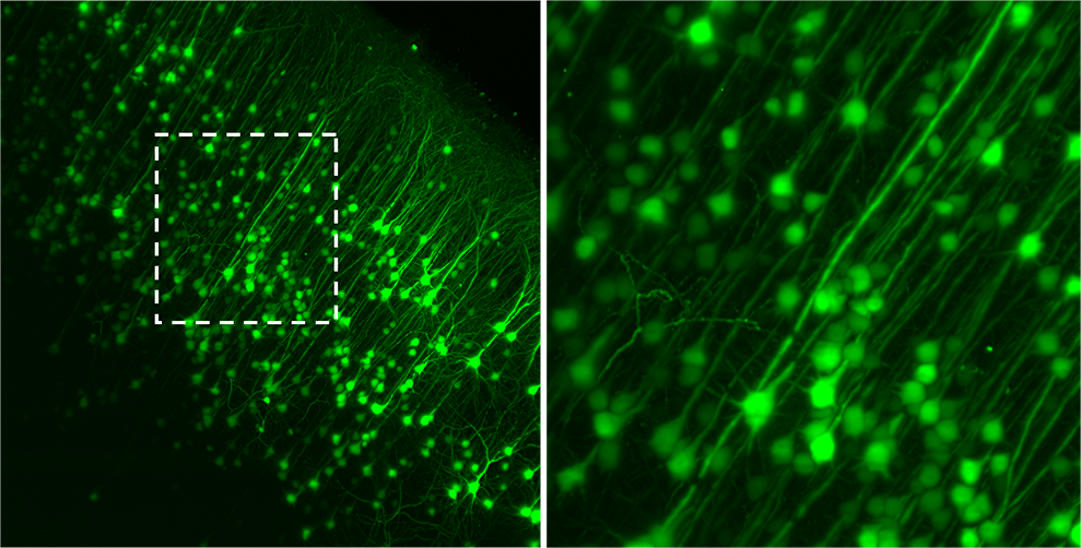
GFP+ motor cortical neurons projecting to striatum via rabies virus expressing eGFP, imaged with SmartSPIM. Sample courtesy of Dr. Byungkook Lim, UCSD.
Upon receiving your samples, LifeCanvas technicians:
You send us: PFA-fixed samples from your experimental animals
Based on your needs, we can provide:

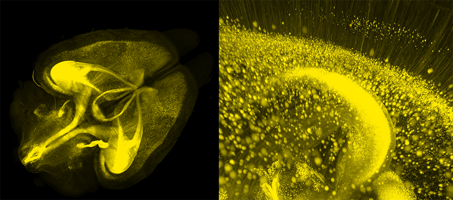
Endogenous Thy1-driven YFP expression in an adult mouse brain, imaged with SmartSPIM. Sample courtesy of Dr. G. Allan Johnson, Duke Center for In Vivo Microscopy.
Here, we demonstrate how the Megatome Premium can easily facilitate this process through whole-mount and non-destructive tissue sectioning.
Immunohistochemistry (IHC) and 3D histology are different approaches to detecting specific proteins in biological tissue.
Tissue clearing techniques have long since sought to maximize optical transparency while preserving sample architecture and minimizing processing time. While...
LifeCanvas tissue clearing is fast, reliable, and cost-effective, allowing uniform and simultaneous clearing for multiple samples.