Immunohistochemistry (IHC) and 3D histology are different approaches to detecting specific proteins in biological tissue.
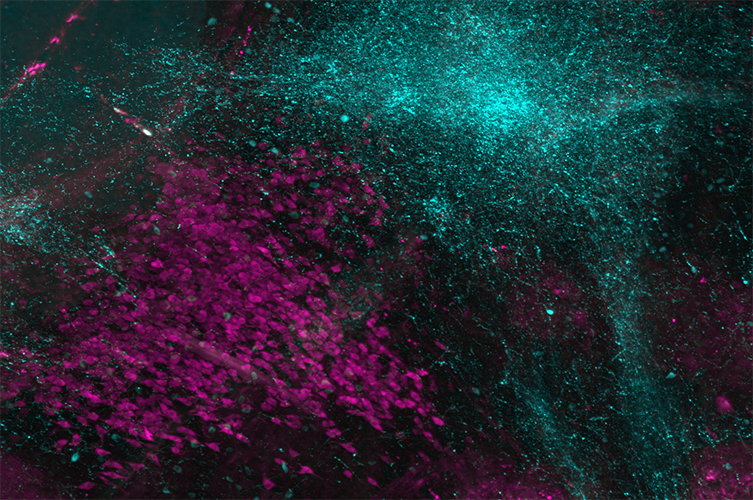
Using our 3D histology platforms, we offer researchers the capacity for whole-organ molecular phenotyping to derive unbiased and illuminating data. Our versatile methods can be employed to map multiple targets, including specific cell types (e.g. neurons, glia), nuclei, and vasculature. We can also process various organ or sample types using tissue-specific markers.
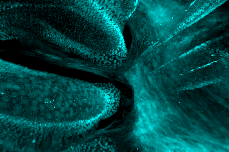
Pictured above: Mouse duodenum perfused with lectin (red) and immunolabeled with anti-Olfm4 (cyan). Sample courtesy of Dr. Suhail Chaudhry, Ferrara Bone Marrow Transplantation Lab, Icahn School of Medicine.


Upon receiving your samples, LifeCanvas technicians:
Based on your needs, we can provide:
Immunohistochemistry (IHC) and 3D histology are different approaches to detecting specific proteins in biological tissue.
What makes the mouse model so popular for disease research? We discuss the benefits, complications, and ongoing improvements of murine...
Mapping human brain organoids in 3D provides neuroscientists with valuable new insights on human brain development and function.
Recent studies leverage LifeCanvas tissue clearing to closely investigate COVID-19's impact on lung tissues.