Acetylcholine neurotransmission in the brain via the central cholinergic system has been implicated in critical functions including cognition, arousal, attention and learning. Moreover, interest in central cholinergic transmission has been piqued given that loss of cholinergic innervation may play an important role in neurodegenerative disorders, particularly dementias. Hence, drugs that elevate acetylcholine transmission in the central nervous system may have the potential to act as memory enhancers and to alleviate cognitive decline.
Cholinergic projections are extensive throughout the mammalian central nervous system emanating predominantly from bundles of neurons in either the basal forebrain or brainstem complexes. Cholinergic neurons of the basal forebrain complex include the medial septum, nucleus Basalis of Meynert and substantia innominate that project to the cortex, hippocampus, thalamus, and olfactory bulb. The brainstem complex (laterodorsal tegmental and pedunculopontine tegmental nucleus) projects to the forebrain, thalamus, hypothalamus and hindbrain. Activation of cholinergic neurons drives arousal, attention and transitions from sleep to wakefulness via the release of the neurotransmitter acetylcholine that binds and activates ionotropic nicotinic and metabotropic muscarinic acetylcholine receptors widely distributed throughout the mammalian central nervous system.
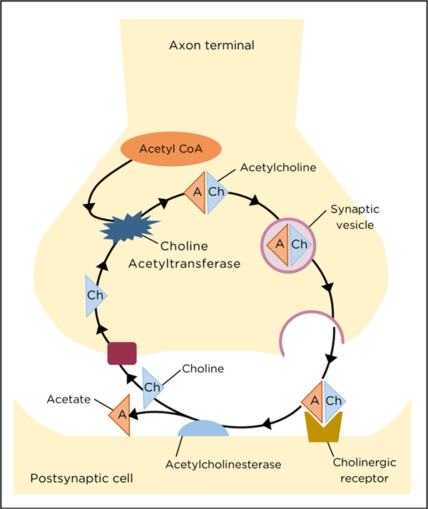
After a neurotransmission event, acetylcholine is degraded in the synaptic cleft by acetylcholinesterase to release acetate and choline which is then retrieved presynaptically via reuptake. Within the presynaptic terminal acetyl-CoA (derived from mitochondria) recombines with choline to produce acetylcholine, a reaction catalyzed by choline acetyltransferase (ChAT). The synthesized neurotransmitter acetylcholine is then repackaged into presynaptic vesicles in preparation for a subsequent transmission event (figure at right).
Since ChAT is critical for the biosynthesis of acetylcholine, it is unique to cholinergic neurons and has been targeted as a biomarker of these neurons within the nervous system. ChAT is produced in the neuronal cell soma and is then transported via axons to the presynaptic terminals to catalyze production of this critical neurotransmitter.
Through LifeCanvas Technologies SHIELD fixing, SmartClear II Pro tissue clearing and SmartLabel immunostaining of whole brain tissues, our SmartSPIM lightsheet microscope reveals the 3D localization of ChAT and highlights cholinergic neurons in the basal forebrain and brainstem complexes. The bundles of cholinergic neurons in these complexes and their projections are clearly visible in the video of coronal and sagittal views throughout the whole mouse brain. Characterization of cholinergic pathways is key to understanding how this important neurotransmitter system impacts brain function in health and disease.
Whole mouse brain processed with LifeCanvas’ tissue processing pipeline. Labelled with anti-ChAT, imaged at 4 µm z-step and 1.8 µm pixel size. Tissue courtesy of the Fishell Lab at the Broad Institute.