At LifeCanvas Technologies, we are driven by a mission to inspire and support cutting-edge scientific discovery. One of our most exciting collaborations is with Dr. Paula Montero Llopis, the Director of the MicRoN Core at Harvard Medical School, who brings her expertise and commitment to empowering researchers through rigorous and reproducible microscopy practices. In this interview, Dr. Montero Llopis shares her insights on the MicRoN Core’s vision, its adoption of LifeCanvas technologies, and how our values align to support scientific advancement.
LC: What is your role as the Director of the MicRoN Core at Harvard Medical School (HMS)?
Paula Montero Llopis: As the Core Director, my responsibilities include managing the facility and its staff, setting our mission statement, providing expertise in experimental set up, training and education in light microscopy, and working with our steering committee that helps guide our core to ensure we meet the needs of our scientific community. My team and I collaborate closely with researchers and industry partners to address their imaging needs. MicRoN operates as a “catalyst core” emphasizing this partnership between core staff, researchers and industry partners in driving scientific discoveries. As a catalyst core, our mission is to educate and empower our users leading to enhanced rigor and reproducibility. This is why LifeCanvas resonated with me—the exceptional support your team provides throughout the imaging process, along with your expertise in sample preparation, imaging, and data analysis, aligns perfectly with our mission.
"MicRoN operates as a “catalyst core” emphasizing this partnership between core staff, researchers and industry partners in driving scientific discoveries. As a catalyst core, our mission is to educate and empower our users leading to enhanced rigor and reproducibility. This is why LifeCanvas resonated with me—the exceptional support your team provides throughout the imaging process, along with your expertise in sample preparation, imaging, and data analysis, aligns perfectly with our mission."
LC: Can you give an overview of how the MicRoN Core is structured, how it collaborates with researchers across various disciplines, and the types of research areas and tissue samples most commonly worked on by your users?
Paula Montero Llopis: We have a very diverse user base in the core, including researchers focused on bacterial cell biology, infectious disease studies, host-pathogen interactions, developmental biology, neurobiology, cancer immunology, tissue architecture, tool development to investigate human disease and whole organ or organism imaging in both live and optically cleared samples. Our core serves as a platform for collaboration across disciplines, offering imaging resources and expertise to researchers with a wide range of scientific backgrounds. We’ve imaged all sorts of tissues and model organisms—gut, brain, liver, skin, lung, you name it, mostly in sections but now we’re moving toward whole tissue clearing. We’ve been able to support clearing without a light sheet microscope, but the solution was not optimal. Most samples are from mammals, but we’ve also worked with clinical samples from affiliated hospitals and a variety of cell types like immune cells and stable cell lines.
LC: Why is the addition of active tissue clearing and light sheet microscopy through SmartBatch+ and MegaSPIM a valuable advancement for the Light Microscopy Core?
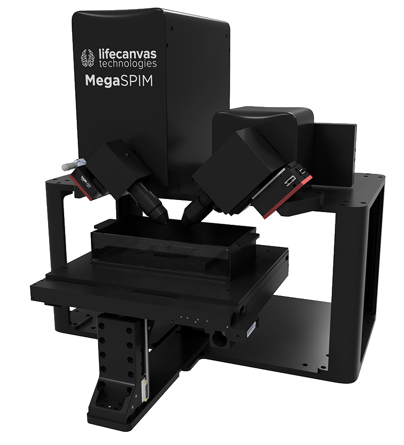
"MegaSPIM addresses this need by offering a versatile and user-friendly solution that works with various clearing methods. Its accessible software and high throughput capacity—allowing us to image multiple samples efficiently—make it especially valuable for our core."
Paula Montero Llopis: Light sheet microscopy was a critical addition for us because we didn’t have a great solution at Harvard Medical School for visualizing large tissue structures in 3D that met the diversity of needs. Tissue clearing is essential for studying the architecture of tissues and understanding cellular relationships. We particularly liked the MegaSPIM as it addresses this need by offering a versatile and user-friendly solution that works with various clearing methods. Its accessible software and high throughput capacity—allowing us to image multiple samples efficiently—make it especially valuable for our diverse user base, including those with limited microscopy experience. We are enjoying collaborating with the LifeCanvas’ team as they also keep us informed about ongoing advancements in their technologies and ensure we have access to the latest improvements.
LC: What current challenges faced by researchers can these technologies address, particularly in terms of efficiency and reproducibility?
Paula Montero Llopis: Our core is very focused on rigorous and reproducible image-based science. We even published a paper in Nature Methods on best practices, which we incorporate into our training sessions. Adopting new technologies can be intimidating for non-microscopists, and that’s where collaborating with LifeCanvas is so valuable. LifeCanvas shares our commitment to high-quality, reproducible science, which gives us confidence that we can provide our users with consistent support. The engineering and support team at LifeCanvas goes above and beyond by not only providing cutting-edge technology but also tailoring it to meet our specific needs, providing invaluable support every step of the way.
"LifeCanvas shares our commitment to high-quality, reproducible science, which gives us confidence that we can provide our users with consistent support. LifeCanvas goes beyond providing cutting-edge technology by customizing and supporting our specific needs, which is invaluable for us."
LC: Based on your hands-on experience with SmartBatch+ and MegaSPIM, what features or capabilities stand out the most, and how do you think they will enhance research outcomes for core users?
Paula Montero Llopis: With the SmartBatch+, we highly value how easy it is to use and its efficiency. The protocols you provide are straightforward, and the batch staining and clearing capabilities make a once painful process much easier and more reproducible. The LifeCanvas team is always willing to work with our trainees to modify the protocols and optimize their specific needs.
For MegaSPIM, I love the straightforward sample mounting—no need for hanging the sample—and its ease of use. It’s especially valuable because you can image several samples quickly and don’t have to set identical parameters for each sample, which is essential when you’re dealing with such diverse needs.
LC: Are there any upcoming projects or research areas you’re especially excited to explore with SmartBatch+ and MegaSPIM technologies?
Paula Montero Llopis: I’ve really enjoyed collaborating with the Wyss Institute at Harvard University and Peter Sorger’s lab, head of the Harvard Program in Therapeutic Science (HiTS) and its Laboratory Systems Pharmacology (LSP) laboratory at Harvard Medical School. They’re focusing on multiplexing with MegaSPIM and will be working on both clearing and multiplexing projects, which I’m thrilled to support. Imaging has been a challenge for many researchers, so having the opportunity to improve their experience and results is incredibly exciting.


