3D labeling & imaging of Neuropeptide Y (NPY) localization
Neurons communicate not only via conventional neurotransmitters but also through neuropeptides. Whereas neurotransmitters often operate by changing the excitability of neighboring neurons, neuropeptides typically impact molecular pathways within target cells leading to diverse modulatory effects. Individual neurons can release both a conventional neurotransmitter and one or more neuropeptides, providing the potential for multiple avenues of signaling.
Neuropeptide Y (NPY), a 36 amino-acid peptide, is an important focus of biomedical research given its abundance in the central and peripheral nervous systems. Furthermore, it is implicated in a wide variety of systems and behaviors including food intake and satiety, mood, neurogenesis, bone growth and cardiovascular regulation. As a result, NPY receptor ligands have been explored as potential therapies for a variety of disorders such as depression, epilepsy, obesity, osteoporosis, and cancer.
Characterizing and comparing the diverse effects of NPY is critical to understand the role of this important neuropeptide and determine its therapeutic efficacy. Through whole-brain imaging NPY-expressing neurons can be identified and examined, enabling the anatomical comparison of NPY pathways simultaneously. LifeCanvas Technologies is at the forefront of whole-brain clearing and imaging techniques and has demonstrated expertise in labeling and imaging NPY within the murine central nervous system. This technology will be essential for advancing NPY-related research and therapies.
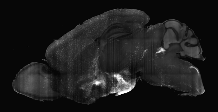
Figure 1: 400-µm MIP of mouse brain right hemisphere stained with Neuropeptide Y.
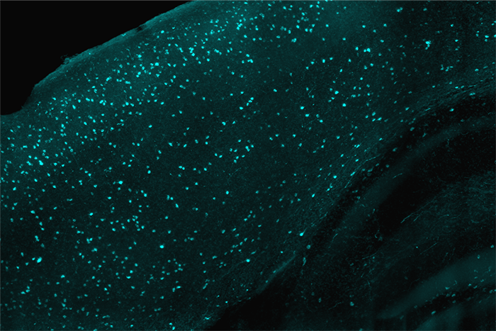
Figure 2: 400-µm MIP of Neuropeptide Y staining in mouse brain dorsal cortex.
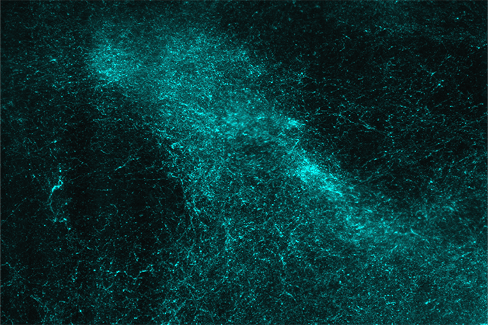
Figure 3: 400-µm MIP of Neuropeptide Y staining in mouse brain ventral region near hypothalamus.