Antibody link: SySy 340 008
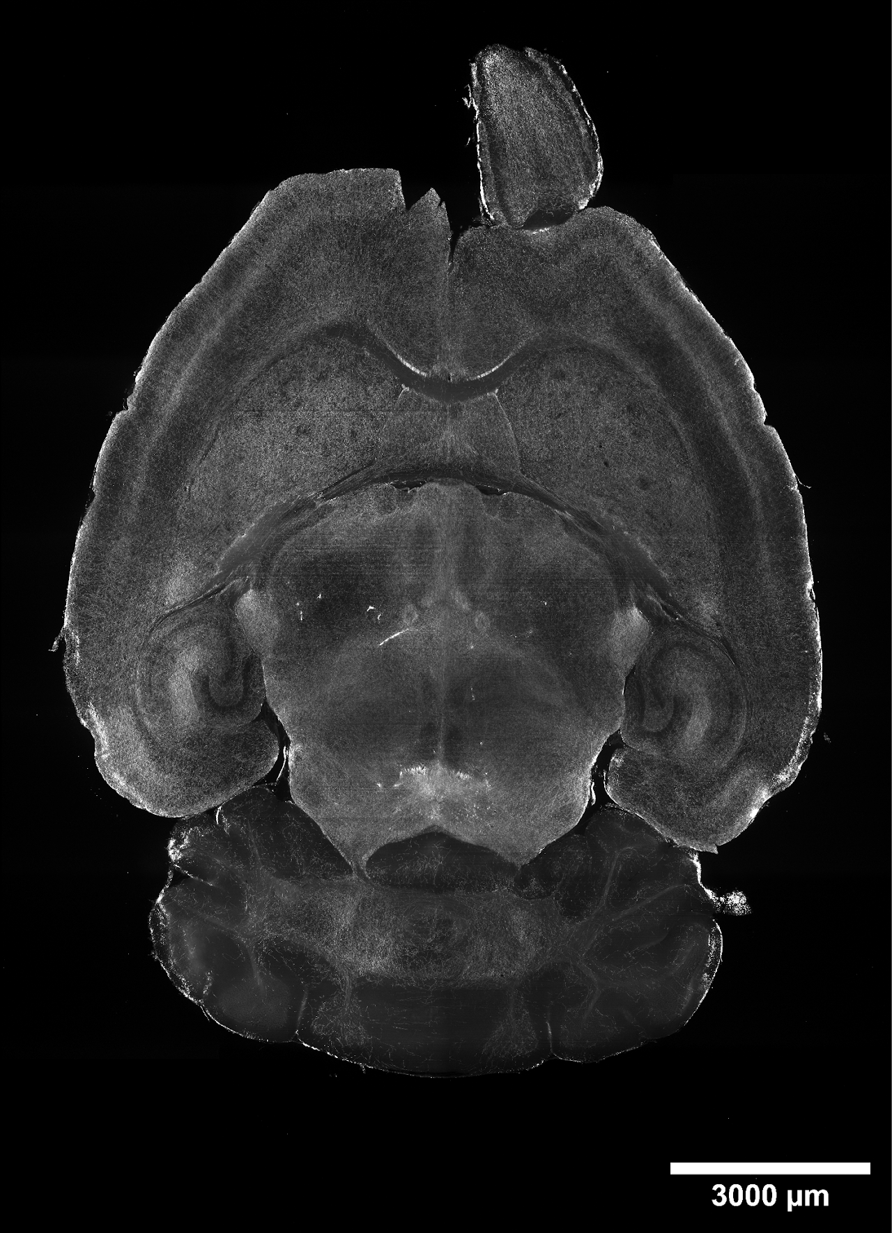
Rabbit Serotonin staining in a whole mouse brain (40 µm max intensity projection).
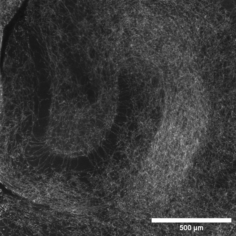
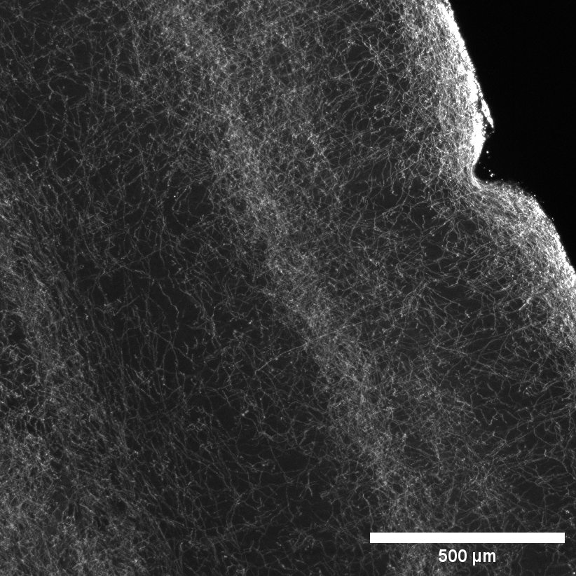
Zoom in of hippocampus (left) and cortex (right) below.
1. Standard SHIELD protocol.
2. Passive delipidation with Clear+ Delipidation Buffer.
3. 2 days blocking with Blocking Buffer at 37°C.
4. Radiant Labeling in SmartBatch+ using 5 µg Rabbit Anti-SERT antibody in the Single Sample Staining Cup.
5. Secondary Labeling with 10 µg Donkey anti-Rabbit SeTau-647 in SmartBatch+.
6. EasyIndex index matching and mounting.
7. Imaged on SmartSPIM with 3.6X objective.