Tatiyana Adkins is the lead author on a recent JNeurosci paper uncovering the neural circuitry behind alcohol drinking and aversive...

Organoids and other complex cell-based models (e.g. spheroids, assembloids) help us capture the heterogeneity of biological tissues in 3D, incorporating multiple cell types and even vasculature. But even as these technologies advance, most high-throughput imaging applications require sectioning.
Why create a 3D tissue, only to lose that spatial context during the imaging process? With the DALISPIM open-top light sheet microscope, you don’t have to. DALISPIM combines the volumetric imaging capabilities of light sheet microscopy with the accessibility of an inverted, open-top configuration.
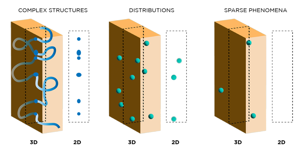
Light sheet fluorescence microscopy (LSFM) is an optical imaging technique used to acquire 3D volumetric datasets. Unlike 2D imaging techniques, LSFM optically sections intact tissues so you don’t have to cut up your sample and lose valuable spatial information.
This figure shows how different phenomena may appear in 3D versus 2D. Continuous structures like vasculature appear fragmented, features that are distributed throughout a tissue depth are not well-represented, and rare features may not be visible at all.
The DALISPIM light sheet microscope allows you to quickly and easily capture 3D data directly from standard sample carriers like multiwell plates. You can use it for high-throughput screening when scale is the priority, or high-resolution data capture from a single organoid when you want a closer look.

Light sheet microscopy doesn’t need to slow down your entire workflow. With DALISPIM, all you need to do is plate your organoids, cover them with EasyIndex matched immersion oil, and snap the whole plate into place. No complex sample prep or mounting steps, and no DIY headaches. When you’re ready for the next plate, simply swap them out and keep moving.
Tatiyana Adkins is the lead author on a recent JNeurosci paper uncovering the neural circuitry behind alcohol drinking and aversive...
We caught up with Dr. Bedenbaugh about her experience as a new investigator, what starting a new lab entails, and...
Dr. Montero Llopis shares her insights on the MicRoN Core’s vision, its adoption of LifeCanvas technologies, and how our values...
Dr. Tanya Daigle used LifeCanvas' services to acquire 3D images of spinal motor neurons illuminated by enhancer AAV.