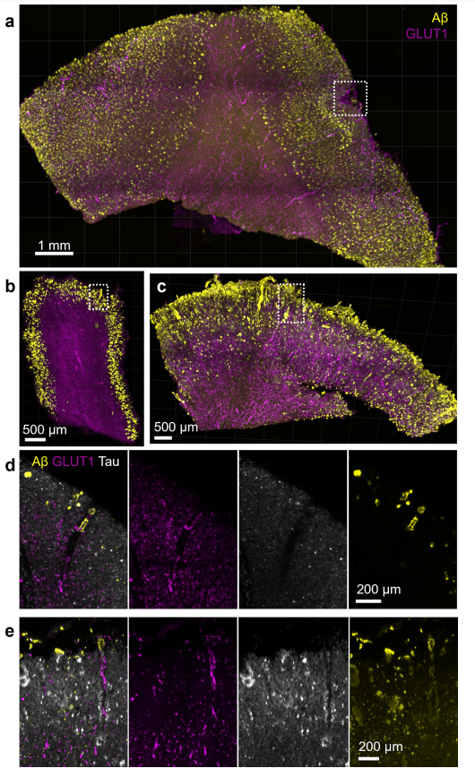
Figure 3 (Ref 5): (a) Tissue from donor AD 6 shows areas with leptomeningeal CAA pathology, (b) tissue from donor AD 5 shows a rare CAA-positive vessel, (c) frequent CAA-positive vessels were observed in donor AD 1, (d) closeup of the dashed box in b, (e) closeup of the dashed box in d.
Movie S1 (Ref 5): CAA pathology and tau accumulation. Article licensed under Creative Commons Attribution 4.0 International License.
For additional publications supporting this use case refer to: