Meet Andrew Stone, the manager of Brandeis University’s Light Microscopy Core Facility, where he’s a hands-on resource for researchers navigating the facility’s 13 advanced microscopes. From training users and advising on optimal imaging tools to collaborating on experimental design, Andrew supports a wide range of projects. Brandeis’ strong legacy in molecular cell biology and neuroscience makes his role pivotal in helping researchers achieve breakthrough insights. Discover how a recent demonstration of LifeCanvas Technologies’ SmartBatch+ and SmartSPIM has convinced Andrew that these systems could be game-changers for the core’s capabilities!
LC: Why is the addition of active tissue clearing and light sheet microscopy a great next step for the Light Microscopy core? What current researcher pain points do they help address?
Andrew Stone: When I joined Brandeis about a year ago, I reviewed our existing instruments and identified some gaps in our offerings. Given the significant amount of neuroscience research and brain slice imaging being conducted, I realized our most pressing need was the ability to perform large-volume imaging. To address this, I began encouraging our team to explore light sheet microscopy, specifically with cleared tissue. With SmartBatch+ we saw the ease of tissue clearing and with SmartSPIM we experienced unparalleled speed in imaging large volumes. Traditionally, researchers here were processing numerous brain slices to gather data, but I wanted to show them that with light sheet technology, they could achieve comprehensive, unbiased imaging of the whole brain in a streamlined, almost hands-off manner. This capability has proven to be incredibly valuable for our work.
LC: Based on your experience with SmartBatch+ and SmartSPIM, what would you like to especially highlight about these technologies and how will they improve data outcomes for core users?
"With SmartBatch+ and SmartSPIM, even people who had never used light sheet microscopy before were able to acquire fantastic data in a very short amount of time."
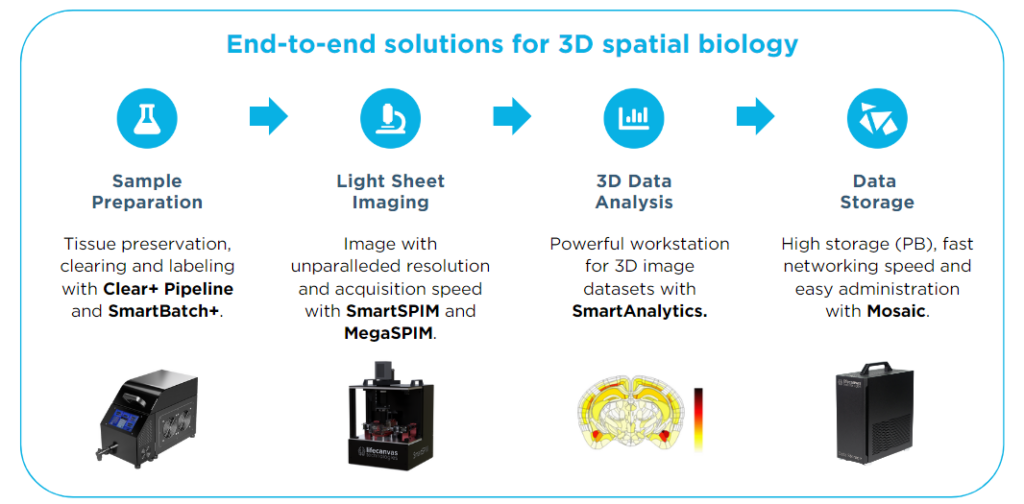
Andrew Stone: Traditional imaging methods, like point scanning or wide-field systems, can be quite time-consuming. While wide-field and spinning disc systems are fast, light sheet imaging takes it to the next level by handling volumes that those other systems just can’t match. Moreover, the ease of use of the systems is great. With SmartBatch+ and SmartSPIM, even people who had never used light sheet microscopy before were able to acquire fantastic data in a very short amount of time. Another advantage is that with LifeCanvas, you get a full end-to-end integrated solution. From tissue clearing to light sheet imaging, making the whole process more streamlined, which is a huge plus.
"With LifeCanvas, you get a full end-to-end integrated solution. From tissue clearing to light sheet imaging, making the whole process more streamlined, which is a huge plus."
LC: How do you foresee current Light Microscopy Core Facility users (utilizing 2-photon, confocal, etc.) utilizing the SmartBatch+ and SmartSPIM to add to their current research data?
Andrew Stone: The SmartSPIM offers a fantastic implementation of axially sweeping light sheet microscopy—it’s highly accessible and efficient. However, one of the biggest barriers to entry for light sheet microscopy has traditionally been tissue clearing, especially for large-volume samples. This is where the SmartBatch+ really shines; it has streamlined the clearing process significantly, particularly for brain tissues, where it has performed flawlessly every time.
I can see some users, especially those currently focused on slice-based imaging, gradually transitioning to using the SmartBatch+ and SmartSPIM. While slice-based methods might still be useful, the speed and hands-off nature of the SmartSPIM system make it a strong alternative. It frees up time and streamlines the entire pipeline from sample preparation to data acquisition and analysis.
I can see some users, especially those currently focused on slice-based imaging, gradually transitioning to using the SmartBatch+ and SmartSPIM.
For tissues where sectioning is complex and reconstruction is difficult, the SmartBatch+ and SmartSPIM open up new capabilities. Even though light sheet microscopy may not achieve the same subcellular resolution as point scanning confocal microscopy with a high NA objective, there’s no need to abandon one method for another. We are looking at complementing the whole-volume imaging with the SmartSPIM by subsequently slicing the tissue to perform higher-resolution imaging on other systems. This dual approach could enhance the overall research workflow for many users.
LC: What upcoming projects or research areas are you excited to explore using SmartBatch+ and SmartSPIM? What people did once they see the capabilities Neuroconnectivity (connections with different parts of the brain) - whole organ visualization.
Andrew Stone: Honestly, since I’m no longer working directly at the bench, I can’t comment on specific projects, but I’m definitely excited to see what others will do with the SmartSPIM once they experience its capabilities. For instance, there are some teams here working on neural connectivity—mapping connections between different parts of the brain. While I’m not a neuroscientist and my language around that field isn’t perfect, I know that studying these connections is much more challenging when using traditional slice-based imaging methods. With the ability to image entire organs and visualize whole volumes, the SmartSPIM could make these kinds of neural connectivity studies significantly more accessible and comprehensive. I would love to see how researchers could leverage the SmartSPIM to push those projects forward, especially in areas where slice-based methods have limitations.


