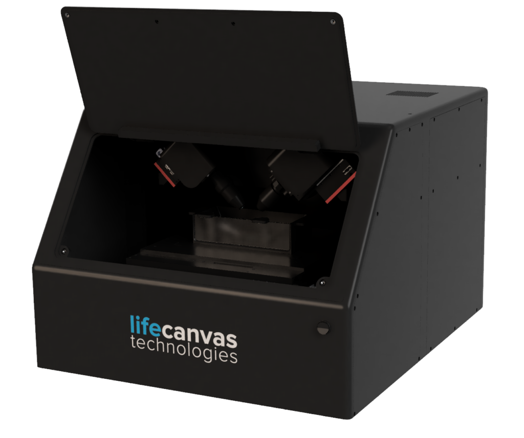
Light sheet fluorescence microscopy (LSFM) is an optical imaging technique used to acquire 3D volumetric datasets from intact or thickly sectioned samples.
Unlike 2D microscopy techniques, LSFM “optically sections” samples by effectively decoupling the paths of illumination and detection into separate light paths, illuminating one z-plane of the sample at a time. This results in volumetric data with minimal photobleaching and low-to-zero background from other z-planes.
Our light sheet microscopes use patented axial sweeping technology (Dean et al. 2015) to speed up imaging time while obtaining high-resolution data across the FOV. An electrical tunable lens, coupled to the back focal plane of a custom-designed illumination objective, generates a light sheet that sweeps axially across the FOV.
The lens is synchronized to the rolling shutter of an sCMOS camera, which captures only the narrowest part of the light sheet. This maintains high resolution across the FOV while omitting out-of-focus regions, enabling rapid image acquisition with uniform axial resolution.
Studying organoids at multiple scales, both in fine detail and in high throughput, is essential to improving their translational relevance. Our DALISPIM microscope gives you the power and flexibility to do just that: capture high-resolution 3D data from multiple organoids at once, directly in a multi-well plate. In this video, acquisition only took 53 seconds per channel!
Light sheet microscopy captures important spatial information often missed by 2D sectioning of human clinical samples, such as heterogeneity, structural complexity, and sparse phenomena. Our DALISPIM microscope makes high-resolution human clinical sample imaging scalable and easy. You can image your samples directly on a slide, dish, or multiwell plate, simply swapping out carriers when you’re ready for your next acquisition.
Video courtesy of Dr. Clarence Yapp, Laboratory of Systems Pharmacology, Harvard Medical School
Mouse and rat models serve a valuable role in studying systemic biological processes, which are inherently spatially complex. SmartSPIM rapidly generates 3D data from intact mouse and rat organs like brain, heart, GI tract, and even whole juvenile mice, giving you spatially unbiased information on structure, connectivity, distribution of phenomena, and more. It’s also compatible with tissues from other small animal models like zebrafish, as well as embryos from various species.
Looking for high-throughput section imaging instead? DALISPIM has you covered.
Most commercial microscopes cannot accomodate tissues from non-human primate (NHP) models, such as marmoset and macaque brains, or require samples to be very thinly sliced. Our MegaSPIM microscope is optimized for high-resolution 3D imaging of large, millimeter-thick samples, maximizing the quality and quantity of data from these tissues.
Biobank specimens are precious and require special care to extract as much information from them in as nondestructive a manner as possible. Most commercial microscopes cannot accomodate the size of these tissues, or require them to be very thinly sliced, losing valuable spatial context. Our MegaSPIM microscope is optimized for high-resolution 3D imaging of large, millimeter-thick samples, maximizing the quality and quantity of data from these tissues.
Visualize neural circuits throughout the brain and even into other parts of the body, like the GI tract and musculature.
Examine intra-brain and brain-body connectivity in 3D using methods like viral tracing (e.g. AAV, rabies), preserving spatial context and continuity.
Get organ-wide insights on how immune cells (e.g. microglia, astrocytes) respond to pathology with high-resolution, spatially-resolved data on interactions between cell types, vasculature, and pathological markers such as amyloid plaques.
Assess the distribution of different therapeutic vectors (e.g. AAVs, LNPs, antibodies) throughout whole organs at cellular resolution. Get quantifiable insights to answer questions like:
Where does my vector localize within the body and within specific organs?
Quantify cells expressing c-fos or other immediate early genes across the brain to investigate regional changes in neural activity without a priori bias. Answer questions like:
Characterize disease-associated pathological markers such as amyloid beta plaques, Lewy bodies, and mHTT aggregates with whole-organ insights, including:
Map blood vessel connectivity and morphology throughout whole organs while preserving valuable spatial context.
Investigate vascular responses to disease states, with detailed 3D information on changes in vessel organization and thickness.
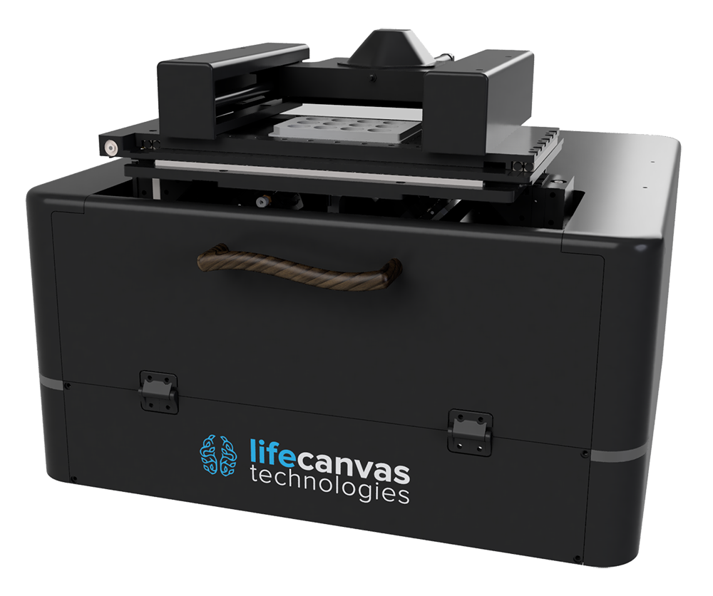
DALISPIM enables direct imaging in standard carriers like multiwell plates, scaling up applications like organoid and clinical sample imaging.
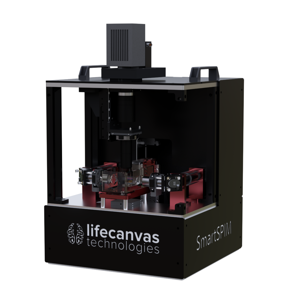
SmartSPIM is ideal for single or sequential imaging of cleared intact tissues like organoids, rodent brains, and marmoset hemispheres.
MegaSPIM is optimized for imaging laterally large samples like millimeter-thick nonhuman primate and human brain slabs and expanded tissues.